


A growth on the hand can be anything from a cyst to a malignant tumor. Use these radiographs to get started on the path towards diagnosis.
A 25 year-old female presented to the emergency department with the complaint of a left hand mass. She first noticed the mass two months prior and it has slowly grown in size. There was no associated pain and she has retained function of her hand. At times she notes a tingling sensation down her index finger. She had no prior hand injury. There has been no drainage from the mass and she denies any fever, chills, nausea, vomiting, night sweats, or unintentional weight loss.
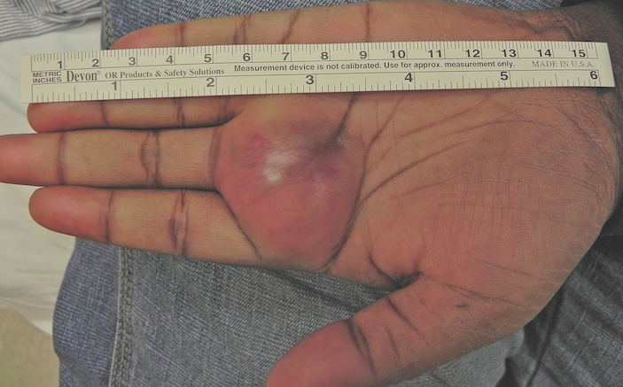
On examination, the patient was in no acute distress, and her vital signs were within normal limits. She had moist mucus membranes. Her neck was soft with no palpable adenopathy. Lungs were clear to auscultation and cardiac exam was normal. The abdomen was soft no tenderness. On the palmar side of the left hand there was a 4×4 cm mass along the MCP joint of the middle finger (figure 1 above). It was soft, immobile, and non-fluctuant. The mass was erythematous and blanching. There was normal finger abduction, adduction, and thumb opposition. The grip strength and wrist flexion and extension were all normal. Sensation of the hand was intact. Radial pulse was intact and there was no thrill or bruit over the mass.

What’s your diagnosis?
- Hemangioma
- Fibroma of the tendon sheath
- Ganglion
- AVM
- Lipoma
- Sarcoma

Radiology
Radiographs of the hand were negative for any fracture or radiopaque foreign body (figure 2 above). There was a focal soft tissue swelling at the base of the 3rd proximal phalanx. An ultrasound was performed and demonstrated a heterogeneous hypoechoic mass measuring approximately 3.5 x 2.5 x 3.3 cm with associated flow just inferior to the 2nd and 3rd digits (figure 3 below).

Discussion
When approaching a hand mass, the first goal is to determine if it is bony or soft tissue in origin. The exam is often telling though a simple x-ray as in this case can distinguish the two. Next, one must determine if mass is infectious, benign, or malignant. A thorough history and exam should exclude infection, as an abscess is fluctuant, warm, and tender and forms over days rather than months. Distinguishing benign from malignant will often require a biopsy. A mass that is large and rapidly growing is more concerning and requires more prompt hand specialist follow-up.

Our patient had a benign presentation and exam. She was referred to an orthopedic hand specialist who established the diagnoses of a fibroma of the tendon sheath after surgical excision. A rare benign soft tissue tumor that more commonly affects males, these lesions are most often found on the hands or fingers [1]. They represent 2-3% of all hand tumors and typically present between the third and fifth decade of life [2]. As with our patient, fibromas are typically slow growing, smooth and well circumscribed [1]. They can cause motor impairment or paresthesias due to their large size and compression of adjacent tendons and nerves. Treatment requires excision that can result in removal of adjacent structures, which has the potential to cause permanent hand dysfunction [2]. Recurrence is rare and there is no risk of malignant degeneration [2].
Other considerations in our case included a giant cell tumor of the tendon sheath and sarcoma, both of which would appear similar on exam and ultrasound to the fibroma of the tendon sheath. MRI can help with the diagnosis though biopsy is required for differentiation. This is an important distinction as giant cell tumors can be more aggressive and sarcomas pose metastatic potential [1].
The most common cause of hand mass and a diagnosis that should always be in the differential is a ganglion cyst [3]. These typically present on the dorsal wrist but they can also present on the volar side of the MCP joint. Classically they are smaller in size and painless though occasionally they can be painful. Patients often note fluctuating size due to a connection of the cyst and joint capsule. On exam, light will pass through the mass and ultrasound will show no internal flow [3]. They are benign and require resection only if symptomatic.

Lipomas are usually diagnosed clinically as they are soft, rubbery, and mobile to palpation. Ultrasound is also very helpful with sensitivity of 88.1% and specificity of 97.9% [4]. Typically lipomas are hyerechoic with well-defined margins and low color flow [5]. They are mostly benign though there is a small risk of malignant degeneration to a liposarcoma and should be removed if growing rapidly or painful [3].
Hemangiomas and AVM’s may also present as hand masses. These are soft and compressible and may have a thrill and bruit on exam. In both cases, plain radiographs and MRI can show calcifications inside within the low-flow vessels of the mass. Ultrasound is also helpful and readily shows the vascular origin [5]. Treatment options include sclerotherapy, arterial embolization, or surgical excision.
Follow Up
The patient followed up in the orthopedic hand clinic the following week. An outpatient MRI further described the mass and two months later she underwent surgical resection identifying the mass as a fibroma of the tendon sheath (figure 4 below). The patient had a full recovery with no motor or neurological deficits.

REFERENCES

- Heckert R, Beart J, Summers T, et. al. Fibroma of the tendon sheath – a rare hand tumor. Pol Przegl Chir. 2012 Dec;84(12):651-6.
- Millon SJ, Bush DC, Garbes AD: Fibroma of tendon sheath in the hand. J Hand Surg Am 1994; 19(5): 788-93.
- Hsu SC, Hentz VR, Yao JR. Tumours of the hand. Lancet Oncol 2007; 8: 157–6.
- Kuwano Y, Ishizaki K, CT; Watanabe R, Nanko H. Arch Dermatol. 2009;145(7):761-764
- Agarwal A, Prakash M, Gupta P, Tripathy S, Kakkar N, Srinivasan R, Khandelwal N. Soft Tissue Masses of Hand: A Radio-Pathological Correlation. Radiology Research and Practice. 2015: 752054.
1 Comment
Hello,
At least now I know what it really is called… a fribroma tumor. My daughter has neurofribromatosis.
My daughter has the exact type of tumor in the palm of her hand–right in the middle. It started small like dime size now it is a golf ball sitting in her palm of hand right in the middle.
I have been begging her for 10 years now to have it removed but she says its not any trouble. She can still use her hand.
Truth is: She can barely make a fist.
How can I convince her that she needs to have it removed?