


Juggling management challenges when handling an agitated patient.
When trauma rolls into the non-trauma center, it can create a number of management challenges. A lot will be dictated by your hospital, EMS system and transport times. When the patient is in extremis, a lot of decisions become simple. Yet, a lot of patients fall into a grey zone in which dilemmas arise on if and when to intubate, pursue imaging and place chest tubes.
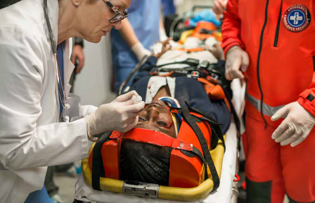
The agitated trauma patient

Agitated trauma patients are a danger to themselves and staff members. While some patients need immediate intubation, there are patients that will respond well to pain medication and/or sedatives. Frequently, trauma patients are under the influence of substances, and haloperidol and/or midazolam administration may prevent an intubation. This is especially helpful in which a neurological exam is crucial to management, such as cases of isolated extremity trauma.
Additionally, some patients with excruciatingly painful injuries such as amputations, deglovings and severe burns may require emergent procedural sedation or delayed sequence intubation.[1] In these patients, consider administration of ketamine 1-2 mg/kg intravenously. This strategy provides excellent pain control, allows for pre-oxygenation, resuscitation and intubation preparation.
Lastly, the agitated patient may be profoundly hypovolemic from blood loss and at risk of a traumatic arrest with premature intubation. Aside from the forced to act intubation scenario, there is usually time to resuscitate prior to intubation. Often, one to two units of PRBCs or whole blood can be administered prior to intubation, as each unit takes ~1 min in a rapid infuser.

Imaging
Do the minimum necessary to facilitate patient care prior to transfer and send all images on a disc along with any reports generated. An e-FAST should be performed on all trauma patients on arrival. The threshold to repeat the e-FAST for a clinical change or prior to transfer should be particularly low, especially for long transfer times. The e-FAST is an additional data point to guide resuscitation. For instance, a patient that is tachycardic, but not hypotensive with a largely positive abdominal FAST may benefit from one to two 1-2 units of blood prior to transfer, especially if EMS is unable to administer blood products en route. The e-FAST is also helpful information for the accepting trauma center team.
It is critical that the actual images, on a disc, are sent with the patient. Without the disc, imaging will be repeated, which exposes the patient to additional radiation and also contributes to potential delays in care. The reports are helpful, as often, radiologists will not generate official reports on outside imaging. Ensuring the transfer of discs and reports is a systems issue, and challenging for an ED physician to navigate while caring for a critical patient. It is highly recommended that there are processes in place for staff to ensure that discs and reports are included in the transfer materials.

Chest tubes, when to place and what size?
Peri-arrest patients with trauma to the chest require expeditious chest tube placement. For more stable patients, proceed with chest x-ray and ultrasound. If both are normal, it is very unlikely the patient needs a chest tube prior to transfer.[2] There is the potential for a pneumothorax or hemothorax to be missed on chest x-ray, but if it’s small, also unlikely they would arrest en route.
Needle thoracostomy can buy time, and it’s important to know if your EMS crew is trained in this technique and what size needles they carry. The recommended needle size is 8 cm, as shorter needles may not enter the chest, especially in muscular or obese patients. For the patient with a significant mechanism, especially if placed on positive pressure, repeat the lung ultrasound prior to transfer.
The plot thickens when there is a delay in the pickup and/or the transfer time is long. Ideally, there should be no delays in transfer to a higher level of care due to imaging. Yet, if the EMS pickup is delayed, a CT may be helpful in some cases. For example, if a patient has a severe blunt mechanism or penetrating injuries to the chest, has a normal chest x-ray on arrival, and gets intubated, the concern is that there could be an occult pneumothorax that will worsen en route.

A CT may show a significant pnemothorax in which a chest tube is beneficial. Sometimes there are patients with very abnormal vital signs — perhaps related to ingestions in the setting of trauma— and it is difficult to determine if they are in shock from a trauma related etiology. A CT may provide additional information that prevents unnecessary blood products or other procedures.
No one needs a 36 Fr chest tube. A 36 Fr chest tube is extremely unpleasant for the patient, and for some smaller patients, it is difficult to place large tubes in between small rib spaces. Most trauma surgeons will recommend 28-32 Fr in cases with potential hemothorax.[3] In the stable trauma patient that is being transferred, it is advisable to reach out to the accepting trauma surgeon for input on tube placement and size, as they ultimately will be managing the sequelae related to the tube.
For air transport, the threshold to place a chest tube should be low. Bradue et al examined this question in a retrospective study from 2014. The average altitude of medical transport helicopters is 2000 ft, which translates into an increase in the volume of closed air space by approx 10%.[4]
In mountainous regions, helicopters will fly higher, which will further increase the risk of pneumothoraces expanding en route. In this study, they reviewed patients that arrived at a trauma center with radiographic evidence of pneumothorax and no chest tube was placed prior to transfer. Of 66 patients included, seven deteriorated in flight. Looking at the cases, a few themes emerged: patients that were intubated, multiple GSWs or other high mechanisms were more likely to develop tension physiology.
Closing transfer advice
Emergency physicians make the best decisions possible at the time with the information and resources available. The potential delays and complications with transfers are numerous. If possible, following up trauma transfers to identify process improvements and increase coordination of care with EMS and trauma centers is an important way to improve trauma transfers. There really is no one size fits all transfer scenarios. Collaboration is needed for all professionals involved along the trauma care continuum.
References:
- Weingart SD, Trueger NS, Wong N, Scofi J, Singh N, Rudolph SS. Delayed Sequence Intubation: A Prospective Observational Study. Ann Emerg Med. 2015 Apr;65(4):349-55.
- Molnar TF. Thoracic Trauma Which Chest Tube When and Where? Thorac Surg Clin. 2017 Feb;27(1):13-23.
- Inaba K, Lustenberger T, Recinos G, Georgiou C, Velmahos GC, Brown C, Salim A, Demetriades D, Rhee P. Does Size Matter? A Prospective Analysis of 28-32 Versus 36-40 French Chest Tube Size in Trauma. J Trauma Acute Care Surg. 2012 Feb;72(2):422-7.
- Braude D, Tutera D, Tawil I, Pirkl G. Air transport of patients with pneumothorax: is tube thoracostomy required before flight?. Air Med J. 2014;33(4):152‐156. doi:10.1016/j.amj.2014.04.009