


Consider patients with known autoimmune conditions before treatment.
A 31-year-old male with a past medical history of ulcerative colitis presented three days after receiving his first Moderna COVID-19 vaccination to the ED with a painful purpuric rash over the right deltoid region beginning two days after vaccination. He reported a raised area and pain over the deltoid region at the site of injection beginning the day afterwards. On Day 2, the purpuric rash developed and spread distally to the anterior arm.
On review of systems, the patient denied fever, chills, cough, congestion, shortness of breath, numbness or weakness. He admitted to pain in his right arm and swelling.

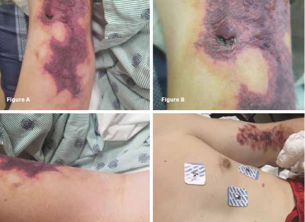
On examination, he was pale but, non-toxic appearing with the following vital signs: T 99.3 F, Pulse 142, Resp 17, BP 113/81, Sat 98% on room air. Lungs were clear. Cardiovascular exam was regular rate and rhythm with intact pulses and perfusion in all extremities. The neurological exam was normal. Skin exam revealed a well demarcated area of purpura, slightly raised with scattered raised fluid-filled blisters, over medial right upper arm. No other bruising, purpura or petechiae on the remainder of his skin. (Figure A, B, C). Musculoskeletal exam revealed profound swelling of medial upper right arm extending through elbow into proximal forearm. ROM of elbow limited somewhat by swelling, but otherwise full range of motion.
Clinical course:
Ultrasound of the right upper extremity demonstrated an occlusive thrombus in the right internal jugular vein and right subclavian vein.

Laboratories were remarkable for the following: WBC 11.56, Hg 10.4 g/dL, Platelet count 854,000, Sodium 129 mmol/L, Potassium 3.8 mmol/L, Bun 4 mg/dL, Creatinine 0.76 mg/dL, Fibrinogen 716 mg/dL, D-Dimer 10.11 ug/dL, CK 355 U/L and SARS-COV2 rapid PCR was positive.
The patient was admitted. Hematology and dermatology were consulted. A heparin drip was started with DVT dosing parameters.
Dermatology saw the patient, diagnosed retiform purpura and performed a biopsy on the lesion. Hematology recommended apixiban as an outpatient. The patient was discharged after a two day hospitalization.

The patient returned to the ED two days later (seven days after vaccination) with a chief complaint of forehead swelling and purpuric rash on his left arm (Figure D). In the ED, a new purpuric rash was discovered on the left arm.
Biopsy results on the right arm were available at that time and demonstrated leukocytoclastic vasculitis in venules and were negative for direct immunofluorescence with epidermal necrosis and blister formation.
The patient was admitted and started on empiric steroids (Methylprednisolone 125mg IV once, then 125mg IV daily. Dermatology was re-consulted and agreed with steroid taper with consideration of colchicine or dapsone if needed in the future, given the working diagnosis of vasculitis.
After undergoing an unremarkable hospital course, the patient was discharged two days later. Patient was negative for concerning results of HIV, Hep C, cryoglobulin, cryofibrinogen, G6PD level (was actually slightly high) testing. The patient’s only ANCA testing that was abnormal was atypical pANCA, which is consistent with his history of UC.

Case discussion:
By definition, vasculitis results from infiltration of vessel walls by white blood cells. The resulting damage generally leads to bleeding and ischemia downstream causing subsequent organ dysfunction, which produce varied clinical signs and symptoms. There are numerous ways to classify vasculitis. This can be done based upon the underlying cause (such as with syphilis or hepatitis C), or by the affected organ (such as with vasculitis limited to the skin), or lastly by the size of the affected vessel.
One of the most commonly used systems for naming vasculitides is from the International Chapel Hill Consensus Conference[1] with the diseases broadly divided by vessel sizes affected. This categorizes vasculitis into large vessel disease (such as Giant Cell arteritis), medium vessel disease (such as Kawasaki) and small vessel disease (such as Henoch-Schonlein Purpura). Initial work up with patients presenting in the emergency department should begin with a CBC with diff (looking for evidence of anemia, leukocytosis or eosinophilia), ESR and CRP (elevation in either suggesting inflammation), cryoglobulinemia panel or HCV testing (pointing most likely to HCV[2]) and consideration of ANCA, and urinalysis (looking for hematuria).
A biopsy may be exceptionally helpful in anchoring the diagnosis, but is ultimately unlikely to change ER management. Consideration for admission versus discharge should be made based upon the patient’s hemostability, the presence or absence of significant systemic findings, as well as the patient’s ability to obtain appropriate care and follow up as an outpatient.
Regarding this patient with Leukocytoclastic vasculitis (LCV), this illness is traditionally grouped under cutaneous small vessel vasculitis[3] (though it can be from systemic vasculitis), and is defined as a combination of neutrophil breakdown and fibrin deposition with damage to vessel walls. LCV does have its own differential diagnosis.[4]
ANCA-associated vasculitis can result in LCV, but the patient’s lack of systemic symptoms in addition to being negative for ANCA makes this less likely. LCV is also associated with immune complex vasculitis, but for this patient, complexes were not seen on the biopsy. UC has been linked to several cases of LCV, [5,6] however LCV is much more commonly linked to RA, SLE and other autoimmune diseases rather than UC.[4]
LCV can also result from an infection, neoplasm or medication, including vaccinations. In terms of infectious causes, strep throat has been linked to causing skin-limited LCV, [4] though this patient did not report any symptoms of such. There have been cases of COVID linked to LCV, [7-9] as well as with the vaccine for COVID[10] though in general there has been insufficient evidence to show a strong causal relationship between vaccines and vasculitis. [11] This case may suggest otherwise, however more research would need to be done to determine causality. More commonly vasculitis is linked to beta-lactams and NSAIDs.[4] Of particular interest in this case is that the lesions started on the arms when typically LCV starts on the legs.
Given the inflammatory nature of LCV, anti-inflammatory agents are the mainstay of treatment. Steroid therapy is typically reserved for treating LCV when lesions are severe — as in this case — or if they are persistent or recurrent. Other pharmacotherapies that have shown efficiency in LCV include dapsone[12] and to a lesser extent colchicines, [13,14] which are typically reserved for recurrent or refractory cases.
Conclusion:
Retiform purpura (Figure A, B, C) is a striking physical exam finding that can be seen in many inflammatory conditions. [15] This patient with ulcerative colitis was vaccinated for COVID-19 while unknowingly being already positive for infection. This likely led to the heightened inflammatory state that precipitated his leukocytoclastic vasculitis and subsequent retiform purpura.
The inflammatory state persisted and worsened until treatment with steroids was finally initiated. Some argument might be made from this case that perhaps individuals with known autoimmune conditions should be tested for COVID-19 prior to vaccination. More investigation into that consideration remains and this warrants further research.
References:
- Jennette, J.C., Falk, R.J., Bacon, P.A., Basu, N., Cid, M.C., Ferrario, F., Flores-Suarez, L.F., Gross, W.L., Guillevin, L., Hagen, E.C., Hoffman, G.S., Jayne, D.R., Kallenberg, C.G.M., Lamprecht, P., Langford, C.A., Luqmani, R.A., Mahr, A.D., Matteson, E.L., Merkel, P.A., Ozen, S., Pusey, C.D., Rasmussen, N., Rees, A.J., Scott, D.G.I., Specks, U., Stone, J.H., Takahashi, K. and Watts, R.A. (2013), 2012 Revised International Chapel Hill Consensus Conference Nomenclature of Vasculitides. Arthritis & Rheumatism, 65: 1-11. https://doi.org/10.1002/art.37715
- Terrier, Benjamina,b,c,d; Cacoub, Patricea,b,c,d Cryoglobulinemia vasculitis, Current Opinion in Rheumatology: January 2013 – Volume 25 – Issue 1 – p 10-18. doi: 10.1097/BOR.0b013e32835b15f7
- Loricera J, Blanco R, Ortiz-Sanjuán F, Hernández JL, Pina T, González-Vela MC, Calvo-Río V, Rueda-Gotor J, Alvarez L, González-López MA, Marcellán M, González-Gay MA. Single-organ cutaneous small-vessel vasculitis according to the 2012 revised International Chapel Hill Consensus Conference Nomenclature of Vasculitides: a study of 60 patients from a series of 766 cutaneous vasculitis cases. Rheumatology (Oxford). 2015 Jan;54(1):77-82. doi: 10.1093/rheumatology/keu295. Epub 2014 Jul 26. PMID: 25065012.
- Fraticelli P, Benfaremo D, Gabrielli A. Diagnosis and management of leukocytoclastic vasculitis. Intern Emerg Med. 2021;16(4):831-841. doi:10.1007/s11739-021-02688-x
- Akbulut S, Ozaslan E, Topal F, Albayrak L, Kayhan B, Efe C. Ulcerative colitis presenting as leukocytoclastic vasculitis of skin. World J Gastroenterol. 2008;14(15):2448-2450. doi:10.3748/wjg.14.2448
- Tripodi Cutrì F, Salerno R, Lo Schiavo A, Gravina AG, Romano M, Ruocco E. Ulcerative colitis associated with leukocytoclastic vasculitis of the skin. Dig Liver Dis. 2009;41(7):e42-e44. doi:10.1016/j.dld.2008.06.018
- Camprodon Gómez M, González-Cruz C, Ferrer B, Barberá MJ. Leucocytoclastic vasculitis in a patient with COVID-19 with positive SARS-CoV-2 PCR in skin biopsy. BMJ Case Rep. 2020;13(10):e238039. Published 2020 Oct 29. doi:10.1136/bcr-2020-238039
- Mayor-Ibarguren A, Feito-Rodriguez M, Quintana Castanedo L, Ruiz-Bravo E, Montero Vega D, Herranz-Pinto P. Cutaneous small vessel vasculitis secondary to COVID-19 infection: a case report. J Eur Acad Dermatol Venereol. 2020;34(10):e541-e542. doi:10.1111/jdv.16670
- Dominguez-Santas M, Diaz-Guimaraens B, Garcia Abellas P, Moreno-Garcia Del Real C, Burgos-Blasco P, Suarez-Valle A. Cutaneous small-vessel vasculitis associated with novel 2019 coronavirus SARS-CoV-2 infection (COVID-19). J Eur Acad Dermatol Venereol. 2020;34(10):e536-e537. doi:10.1111/jdv.16663
- Bostan E, Gulseren D, Gokoz O. New-onset leukocytoclastic vasculitis after COVID-19 vaccine [published online ahead of print, 2021 Jul 9]. Int J Dermatol. 2021;10.1111/ijd.15777. doi:10.1111/ijd.15777
- Bonetto C, Trotta F, Felicetti P, et al. Vasculitis as an adverse event following immunization – Systematic literature review. Vaccine. 2016;34(51):6641-6651. doi:10.1016/j.vaccine.2015.09.026
- Fredenberg MF, Malkinson FD. Sulfone therapy in the treatment of leukocytoclastic vasculitis. Report of three cases. J Am Acad Dermatol. 1987;16(4):772-778. doi:10.1016/s0190-9622(87)70100-5
- Callen JP. Colchicine is effective in controlling chronic cutaneous leukocytoclastic vasculitis. J Am Acad Dermatol. 1985;13(2 Pt 1):193-200. doi:10.1016/s0190-9622(85)70158-2
- Sais G, Vidaller A, Jucglà A, Gallardo F, Peyrí J. Colchicine in the treatment of cutaneous leukocytoclastic vasculitis. Results of a prospective, randomized controlled trial. Arch Dermatol. 1995;131(12):1399-1402.
- Georgesen C, Fox L, Harp J. Retiform Purpura: A diagnostic Approach. Journal of the American Academy of Dermatology. 2019, Vol 82, Issue 4, pages 783-796