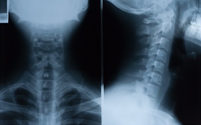
Ultra low-radiation CT technology is sounding the death knell of plain x-rays.
I graduated from medical school in 1970 (shortly after the development of penicillin) so for me the invention of the CT scanner has been, without doubt, the single most important innovation in medical diagnostics.
According to my highly reliable (?) internet sources, the CT scanner was invented in 1972 by Drs. Hounsfield and Cormack. For their extraordinary contribution to medicine they both were awarded the Nobel Prize for Physiology or Medicine in 1979.
CT scanners began being installed between 1974 and 1976 but ordering a CT scan on a patient in the ED was a big deal at that time — you had to get all sorts of consults before pulling the trigger.
Back then it was relatively simple. If a patient had a possible neck injury you either ordered a plain x-ray or you didn’t. As anticipated there were lots and lots of unneeded x-rays (and this was augmented by the fact that every Tom, Dick and Mary who was in the most minor of incidents came into the ED collared and boarded [fortunately that practice is slowly going the way of the dodo]).
To help physicians make more rationale and consistent decisions about who would benefit from a neck x-ray various rules and guidelines were developed. The most famous was the NEXUS decision instrument that was published in the New England Journal in 2000 with the lead author being my close associate, Jerry Hoffman.
Fast forward 20 years and CT scanners are ubiquitous and ordering one is simple – just check the box. Now we have three choices in minor neck injuries – no imaging, plain x-rays or CT scans. Why would anyone who is going to be imaged not get the best imaging? What would you want for yourself or your family? Simple.
And the fact is that unequivocally, CT scans are superior to plain films. So the issues come down to cost and radiation. Can we talk? It’s no secret that CT scanners are printing presses for money. We all know that the astronomical charges for CTs have little relationship to the cost of the study – even including a reasonable profit margin. Here’s some ballpark figures. Medicare will reimburse about $75 for a neck x-ray while it will pay about $150 for a CT of the neck. Charges are another story. Expect to be charged about $300 for a neck x-ray and about $1,700 for a CT – but variance in charges are enormous between facilities. Commercial insurance plans can expect to pay a lot more (generally at least a multiple of what Medicare pays). But, the point is that in reality, CT scans don’t really cost that much – it is the charges that are the problem. And as more and more studies are done, costs per scan should come down (if the laws of economics applied to medicine – but that’s another story)
When it comes to radiation, a plain c-spine x-ray delivers about 0.2 mSv while a CT of the cervical spine delivers about 4-6 mSv (which I find surprisingly high since a head CT is about 2 mSv). The amount of radiation associated with CT scans is very variable based on where and how they are performed. As a result some patients receive a lot more radiation than they need to – and this is especially an issue in young children (who are particularly susceptible to the long-term carcinogenic effects of radiation). To educate the radiologic community regarding what can be done to limit radiation to children the Image Gently project (ImageGently.org) was organized by a consortium of concerned organizations.
The most exciting reports on radiation reduction are coming out of Vancouver General Hospital where, in conjunction with Siemen’s state-of-the-art third generation equipment and low-dose algorithms, radiation doses comparable to those from plain x-rays are being achieved in selected studies. For example, with the Siemens SOMATOM Force dual source scanner (about $3M) images of the chest and abdomen can be acquired in one second. Yes, one second. And what about radiation dosage – how about 0.1 mSv for a lung scan?. I know this sounds like a commercial but the fact is that this is just an example of how fast technology is moving in the CT and MR worlds – and all the major manufacturers are competing for sales of the high-end scanners
So, depending on the equipment being used at your hospital and the care that the radiologists and technicians use in calculating appropriate scanning protocols, concerns about radiation will become less and less of an issue over time.
So, seems the answer is becoming easier – no imaging or imaging with CT. The key is for clinicians to use good judgment in determining just who should be scanned. Unfortunately, just as with plain x-rays, there are way too many scans being done in low-risk patients. Will this ever change? I’m rather pessimistic.
Here are some papers addressing the issues under consideration. The first paper focuses on the patients we are most concerned about – low-risk adults (who represent the vast majority of our cases). The authors note in this four study review that the sensitivity of plain x-rays in the NEXUS trial was 92% and in three other studies of patients who failed the NEXUS criteria (could not be clinically cleared) the sensitivity of plain x-rays was 45%, 65% and 62%. Clearly, very disappointing numbers.
ARE PLAIN RADIOGRAPHS SUFFICIENT TO EXCLUDE CERVICAL SPINE INJURIES IN LOW-RISK ADULTS?
Hunter, B.R., et al, J Emerg Med 46(2):257, February 2014
BACKGROUND: The risk of cervical spine injury in patients with blunt trauma is reported to be between 1% and 3%. Recently the use of clinical protocols and standard three-view radiography to clear the C-spine has been called into question.
METHODS: These multicentered authors, coordinated at Indiana University School of Medicine, assessed the ability of plain x-rays to exclude C-spine injuries in low-risk adult blunt trauma patients. Their literature review identified four studies that addressed this issue.
RESULTS: In the 34,069-patient NEXUS trial, at least one C-spine injury was diagnosed (on the basis of total imaging ordered at the discretion of managing clinicians) in 2.4% of the patients. The sensitivity of “adequate” plain films for C-spine injury was 92.1%. In a second trial of 667 patients who failed the NEXUS criteria (were unable to be clinically cleared), C-spine injuries were diagnosed on the basis of clinical data and CT imaging in 9% of the patients (45% were considered clinically significant), and the sensitivity and specificity of plain x-rays were 45.0% and 97.4%, respectively. The third trial included 1,199 blunt trauma patients who also failed the NEXUS criteria. C-spine injuries were diagnosed in 9.5%. Sensitivities for C-spine injury were 100% for CT scanning but 65% for plain films. The fourth trial included 1,505 blunt trauma patients who also failed the NEXUS criteria, all of whom had three-view plain films and CT scanning. Rates of C-spine injury and “clinically significant” injury were 5.2% and 3.3%, respectively. The sensitivity of imaging for clinically significant injury was 100% for CT scanning, 36% for plain films and 62% for “adequate” plain films.
CONCLUSIONS: The authors acknowledge the methodologic limitations of the reviewed studies, but conclude that the sensitivity of three-view plain radiographs appears to be inadequate to exclude C-spine injury in adult low-risk blunt trauma patients. 18 references
And here’s a paper addressing the work being performed at Vancouver General. It demonstrates that with a combination of specific imaging techniques coupled with the use of a third generation scanners that radiation levels can be markedly reduced and, in some cases, are lower than those with plain x-rays. The authors note that a c-spine CT was obtained at an effective dose of 0.6 mSv compared to 6 mSv in the past. Some of the radiation levels noted in this discussion are truly remarkable.
COMPUTED TOMOGRAPHY AND THE IMPENDING OBSOLESCENCE OF THE PLAIN RADIOGRAPH?
McLaughlin, P.D., et al, Can Assoc Radiol J 64(4):314, November 2013
Exposure to ionizing radiation is generally considered to be a major limitation to widespread use of CT scanning. The authors, from Vancouver General Hospital in Canada, discuss technical breakthroughs that have combined to allow significant CT dose reduction without the loss of image quality. These include automated exposure control, iterative reconstruction algorithms and the use of third-generation CT detectors. The authors provide examples of ultra-low-dose CT scans with information on current and past (2008) radiation dose levels. An ultra-low-dose CT of the cervical spine in a 28-year-old pregnant female was performed at an effective dose of 0.6mSv compared with 6mSV at 2008 levels (and 0.2mSv for the average C-spine x-ray in 2008). The effective radiation dose for an ultra-low-dose chest CT was 0.52mSv, compared with 7mSv for a standard CT and 0.1mSv for an average chest x-ray performed in 2008. The effective radiation dose for an ultra-low-dose coronary CT angiogram was 1mSv compared with average doses of 16mSv and 7mSv for an average coronary angiography and CT of the chest performed in 2008. An ultra-low-dose CT performed to visualize a renal calculus had an effective radiation dose of 0.4mSv compared with average doses of 0.7mSv for an abdominal x-ray and 8mSv for a CT of the abdomen in 2008. Finally, an ultra-low-dose abdominal CT for suspected appendicitis was associated with an effective radiation dose of 0.56mSv. The authors feel that the use of a combination of dose-reducing CT technologies decreases the radiation exposure inherent to CT scanning to levels that are comparable to those of plain films. 16 references
So, is the plain x-ray of the cervical spine dead. Not just yet. But as we effectively deal with radiation and costs there will be no question that physicians will even more preferentially order CTs of the neck than they do now.
But, we have to be realistic. For most of us, we don’t have access to a third generation scanner, but that doesn’t mean our radiologists and techs can’t be more focused on reducing radiation through better protocols. And costs? Well, as more and more care is based on payment models other than fee-for-service, the costs / charges for a CT will become progressively less of an issue and the focus will increase on the improved throughput and image quality advantages that CT scanning provides.


