A 37-year-old man presented to the emergency department with severe respiratory distress. He reported a history of hypertension, diabetes and end-stage renal failure for which he received hemodialysis three times per week. However, he admitted to missing his last two dialysis sessions. The patient was diaphoretic and in obvious respiratory distress. His vital signs were notable for a respiratory rate of 32/minute, pulse oximetry of 80% on room air, heart rate 70/minute, blood pressure 190/100, afebrile, and his fingerstick glucose was 200. He had obvious jugular venous distension and rales on lung auscultation. The rest of his exam was unremarkable.
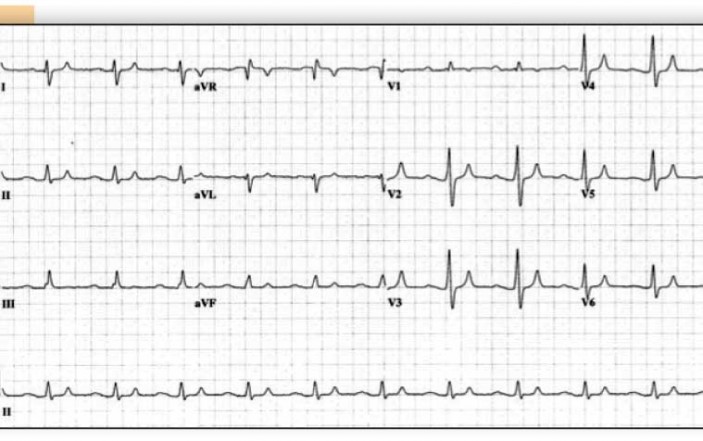
The patient was placed on a cardiac monitor, which demonstrated sinus rhythm with a rate of approximately 70/minute. Intravenous access was obtained and multiple laboratory studies were sent. Oxygen was administered via 100% non-rebreather face mask. An ECG was obtained (see Figure 1) and interpreted as sinus rhythm and an incomplete right bundle branch block pattern. No other abnormalities were noted by the treating physician. A chest x-ray was obtained as well and demonstrated cardiomegaly with pulmonary edema.
 The supplemental oxygen via face mask improved the patient’s pulse oximetry to 93%, but the patient appeared to be fatiguing rapidly. The decision was then made to intubate the patient. Intravenous etomidate and succinylcholine were administered and rapid sequence intubation was successfully performed. The pulse oximetry improved to 100%. However, soon after the intubation, the patient’s cardiac rhythm changed (see Figure 2). The physician caring for the patient noted the regular, wide-QRS complex tachycardia with a rate of 105 beats/min, the absence of obvious atrial activity and the monitor interpretation of “V-TACH.” The patient’s blood pressure remained elevated, and so he diagnosed the rhythm as stable ventricular tachycardia (VT). A 1.5 mg/kg bolus of lidocaine was administered intravenously, and approximately two minutes later, the patient developed asystole (see Figure 3). Standard Advanced Cardiac Life Support protocol was initiated, including intravenous calcium. Unfortunately, the patient never regained pulses. Shortly after the patient was pronounced dead, the laboratory called to report that the initial serum potassium level (prior to intubation) was 7.3 mEq/L.

Discussion:
Several important teaching points can be found in this case. First, this patient’s history of missing two dialysis sessions should immediately prompt concern for hyperkalemia. The initial ECG (Figure 1) is also suggestive of hyperkalemia: slightly peaked T-waves, slight widening of the QRS complexes, rightward axis (large S-wave in lead I) and incomplete right bundle branch block. Hyperkalemia is known to produce almost any finding on the ECG (1), which is one of the reasons that I often refer to hyperkalemia as “the syphilis of electrocardiography” (i.e., the great imitator!). The physician in this case, however, did not notice the slightly peaked T-waves and was primarily focused on managing patient’s overt respiratory distress. Remember that patients with chronic renal failure often “tolerate” much higher serum potassium levels than patients with normal renal function. A serum potassium level of 7.3 mEq/L in a normal patient would likely produce a profoundly abnormal ECG. Yet, in this patient produced more subtle abnormalities.
The second key teaching point or reminder in this case is to be wary of using succinylcholine in patients with known or suspected hyperkalemia. Succinylcholine produces only mild elevations in serum potassium levels in normal patients; but patients with pre-existent hyperkalemia are at risk for developing more dangerous elevations in serum potassium levels after the administration of succinylcholine. For this reason, non-depolarizing paralytics such as rocuronium are preferred in such patients.
The third key teaching point in this case is less well-known than the above points: the diagnosis of VT should be avoided in patients with heart rates < 120-130/min (2-4). In this case, although the post-intubation rhythm (Figure 2) looked like VT, a rate of 105 beat/min virtually excludes this diagnosis. The only exception would be in a patient already being treated with ventricular antidysrhythmics such as amiodarone, in which VT might occur at slower rates. In contrast, there are a few classic conditions that are notorious for mimicking VT but with slower rates: hyperkalemia (2,4,5), sodium-channel blocking medication toxicities (e.g., tricyclic antidepressants, cocaine) (2,4,6) and post-myocardial infarction (MI) reperfusion arrhythmias (2-4,7) are the most notable.
Common teaching in medicine is that when faced with a wide-complex regular tachycardia, the treating physician should always treat the patient for VT. While this teaching is generally true, the use of traditional “VT medications,” such as lidocaine, procainamide or amiodarone, for any of the mimics noted above can actually be deadly. Recall that lidocaine and procainamide are Class I antiarrhythmics—sodium channel blockers. Even amiodarone, which is primarily a Class III antiarrhythmic, does have Class I effects as well. The use of Class I antiarrhythmics for patients with hyperkalemia (which itself is known to poison the sodium channels (5)) or sodium-channel blocker toxicity can produce such pronounced sodium-channel blockade that asystole may result (4-6), such as in this case.
Post-MI reperfusion arrhythmias often take the form of a regular, wide-QRS complex dysrhythmia with a rate of 90-120. The rhythm is often mistaken for VT. Treatment with “standard” VT medications is well-known to induce asystole (2-4,7). Figure 4 is the ECG from a patient in the coronary care unit with an acute MI, approximately two hours after receiving thrombolytics, who developed this regular, wide-QRS complex rhythm with a rate of 115/min . Despite the rate < 120-130/min, the diagnosis of VT was made and the patient was treated with amiodarone. Asystole resulted, and the patient died. Proper treatment of this reperfusion arrhythmia is simple observation; the rhythm is usually transient, lasting no more than a few minutes, and it is not hemodynamically-compromising.

For more teaching cases in electrocardiography, check out ECGs for the Emergency Physician, Volumes 1 and 2 by Amal Mattu and William Brady (Blackwell Publishing).
References:
1. Mattu A, Brady WJ, Robinson DA. Electrocardiographic manifestations of hyperkalemia. Am J Emerg Med 2000;18:721-9.
2. Kastor JA. Arrhythmias, 2nd ed. Philadelphia, PA, W.B. Saunders Company, 2000.
3. Chou T-C, Knilans TK. Electrocardiography in Clinical Practice, 4th ed. Philadelphia, PA, W.B. Saunders Company, 1996.
4. Marriott HJL. Pearls & Pitfalls in Electrocardiography, 2nd ed. Baltimore, MD, Williams & Wilkins, 1998.
5. McLean SA, Paul ID, Spector PS. Lidocaine-induced conduction disturbance in patients with systemic hyperkalemia. Ann Emerg Med 2000;36:615-27.
6. Wang RY. pH-dependent cocaine-induced cardiotoxicity. Am J Emerg Med 1999;17:364-9.
7. Cairns CB, Paradis NA. Empiric lidocaine: déjà vu (all over again?). Ann Emerg Med 2000;36:626-7.


1 Comment
[b]Yes I agree with the comments and teaching given in the above mentioned case. The ECG was for sure an eye opener for the Freshers IN EM , I have seen Lots of time people giving Succs to CKD patients which can increase the Serum K levels for atleast 0.5 , and this could be a disaster for the DOC and the patient on that vary day. Thanks for the VT revision![/b]