EMS brings you a 54 year old who slipped off a wet ladder rung and fell about 15 feet. He is boarded and collared by the paramedics and complains of R hip pain. Vitals are OK, and he complains of pain in his R hip area. “I can stand,” he tells you as you finish your unremarkable primary survey, “but it just hurts . . . a lot”. By exam, he has poorly localizing pain in the R hip and pelvis area, with some pain in the R hip with ROM and axial load. There is no shortening of the R leg, and the N/V exam is normal. You get a quick portable X-ray of the hip, and order the full Monty – total body CT scan – for mechanism of injury
The first portable X-ray is shown (below). What does it show? What else might be helpful?

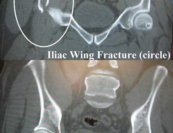
Dx: Iliac Wing Fracture
This is a great x-ray case. Basically, I don’t think you can see this fracture on the first (portable) film. However, the bone windows of the pelvic CT scan show a nice iliac wing fracture, complete with comminution. Ouch. No wonder it hurts to walk. This is also a good example showing that a long-spine board can make seeing a tough fracture even more difficult. Most patients are better served to be log-rolled off this device shortly after arrival in the ED.
This fracture was managed conservatively by ortho. Most major pelvic fractures and those involving the acetabulum will be sent to the referral center by community hospital orthopedists, but this was truly an isolated injury and appeared to have a low risk of associated GU and hemorrhagic complications. The mechanism was presumed to be a direct blow to the lateral aspect of the pelvis, rather than an axial load through the leg and hip joint. In the latter case, one would also look for calcaneus and lumbar spine fractures.
Long Spine Board Immobilization – Roll ‘em off in the ED!
Pre-hospital utilization of a hard spine board (aka “long spine board”) is commonplace. The device is practical for extrication and immobilization of injured or obese patients. However, there is a growing consensus among ED providers that patients be removed from the long spine board ASAP and transferred to a flat-supine position in a hospital bed. Adverse effects of the long spine board include pressure sore development, inadequacies of spinal immobilisation and support, pain and discomfort, respiratory compromise and diminished quality of radiological imaging (see D Vickery, Emerg Med J 2001;18:51-54).
Just as with cervical collars, long spine boards may not really immobilize the axial skeleton all that well, and are probably inferior to devices such as the pre-hospital vacuum matress for that purpose. Rarely we will leave a patient strapped to the board for a brief period – thrashing post-head injury and pre-RSI, or massive obesity in immediate danger of falling off stretcher – but mostly we just log-roll them into the flat supine position. In short, the long spine board is a useful “transport device” for EMS, but really has no role for the hospitalized patient. (See additional related discussion on connect.jems.com/forum/topics/backboards-vs-vacuum-mattress)


2 Comments
Great case. Agree, the board does nothing to protect the spine. At places where they use the board for transport, we’ve used a gel pad that we put between patient and the board when we roll and expose them. Then they get the CT still on the board, but without the pressure ulcer risk.
I am not sure it would have helped but my studies on this patient would have been portable pelvis or at least a kub that opens to show the whole pelvis. Might have helped.