
Injuries to the wrist and carpal bones can seem confusing, but with an understanding of the anatomy you can avoid critical ramifications.
Wrist injuries may be among the most unfamiliar and commonly missed orthopedic injuries among emergency physicians. The anatomy alone is daunting. The injuries can be subtle and when missed can have debilitating repercussions. Many carpal injuries are missed because they are associated with a concurrent fracture pattern. Studies suggest between seven and twenty percent of all distal radius fractures will have an accompanying carpal injury. Every carpal bone can be fractured and I will not discuss each in detail, but instead will focus on the most common or most often missed injuries.
Anatomy
Bones
“Some Lovers Try Positions That They Cannot Handle.” This mnemonic has always worked well for me but it’s dependent on knowing which direction you are labeling the bones. It goes radial to ulnar along the bottom row (scaphoid, lunate triquetrum, pisiform) then radial to ulnar along the top row (trapezium, trapezoid, capitate, hamate). (See figure A below)


Articulations and Ligamentous Stability
The surface of the distal radius has a notch or ridge that separates articulations with the scaphoid and lunate bones. There are a series of ligaments which stabilize the arrangement of the carpal bones. Without these, the small round carpal bones are no more than a bag of marbles. It’s not necessary for the ED physician to know all the carpal ligaments but it is important to recognize patterns of ligamentous disruptions and instability which I will discuss. The two most important ligamentous structures are the scapho-lunate ligament and the luno-triquetral ligament. There are many other stabilizing carpal ligaments but I will focus on these as they relate to very specific injury patterns.
Vascular Anatomy
The vascular anatomy of the hand consists of the deep and superficial palmar arches which are typically damaged in puncture, laceration and crush injuries. Vascular anatomy is uniquely pertinent as it pertains to the scaphoid and the lunate. The scaphoid receives its blood supply from both the dorsal and volar branches of the radial artery with the dorsal responsible for 80% of the blood supply including the proximal half of the scaphoid. The waist separates the proximal and distal pole. This watershed area in the waist complicates bone healing and increases risk of non-union.
Anatomy Pearls

- Healing is dependent on location. Because of the dominant dorsal supply, the further a fracture is from this vascular source the higher the risk of non-Union. The dorsal branch enters on the distal pole, so nearly all distal pole fractures will heal, while up to 83% of proximal pole fractures will result in non-union. The lunate also receives blood supply from both the solar and dorsal surface however twenty percent of lunates only have a volar blood supply. (See figure B below)
- The variability in blood supply to the lunate can influence the likelihood of “Kienbocks Disease” which is avascular necrosis of the lunate.
- The lunate is absolutely key to the stability of the entire wrist because it strongly binds both the scaphoid and the triquetrum which articulate with the distal radius
- Disruptions in the luno-triquetral ligament and scapho-lunate ligament cause wrist instability.
- The vascular supply to both the scaphoid and the lunate can increase risk of nonunion or avascular necrosis.

Scaphoid Injuries
Exam
Scaphoid fractures make up anywhere between fifty and eighty percent of all carpal injuries. Most injuries result from falls on a dorsiflexed hand. There are a number of provocative tests such as the “shift test” and the watson test, but for emergency physicians the two most sensitive and reproducible tests are painful palpation of the scaphoid and pain with axial load of the thumb. Axial load is also called The Grind Test and is best done with the wrist in slight ulnar deviation.
X-ray
Scaphoid fractures may not be evident or recognized on plain X-ray. Because of the vascular anatomy and the risk of non-union, anything short of perfect alignment in proximal pole fractures will result in non-union.
Treatment
Some surgeons operate on all proximal pole fractures, but certainly anything greater than 1mm of displacement is considered high risk. Distal pole injuries will accept more displacement. The threshold for surgical intervention in proximal pole injuries is variable.

Treatment of scaphoid fractures has radically changed in the past few decades. Many hand surgeons suggest, with good evidence, that nearly all waist and proximal scaphoid fractures are operative. Because the operative techniques for fixation have significantly improved, the frequency of operative interventions has significantly increased.
Because many scaphoid fractures are not radiographically visible, any scaphoid pain warrants immobilization. Typically, immobilization is for one to two weeks at which time the patient is re-examined by the hand surgeon. If the pain has completely resolved and follow up X-Ray is normal immobilization is discontinued. If pain persists MRI is indicated.
Because between seven and up to twenty percent of distal radius fractures may have associated carpal injuries, careful attention should be given to the scaphoid during exam and X-Ray evaluation. Any scaphoid pain in the setting of a distal radius fracture warrants a thumb-spica extension to a sugar tong splint.
Pearls
- Check scaphoid tenderness with palpation and axial load.
- Check scaphoid tenderness in distal radius fractures.
- Inform patients of possible of scaphoid fracture with negative X-ray.
- Proximal scaphoid fractures have much lower healing rates than distal fractures.
- Inform patients with obviously displaced scaphoid fractures, or any distal pole fractures, of the high likelihood for operative intervention.
- In all suspected scaphoid injuries emphasize the importance of close follow up with hand surgery
Scapho-Lunate Dissociations
These ligamentous injuries are probably the most frequently missed wrist injuries in the emergency department. The injury results from the same mechanism as distal radius and scaphoid injuries with the exception that there may be additional ulnar deviation at the time of fall.
Exam
Exam is much more subtle and the physician must have clinical suspicion to recognize the findings. The scaphoid itself may not be painful but the patient will have pain or swelling over the dorsal aspect of the wrist, particularly over the scapho-lunate space.
X-ray
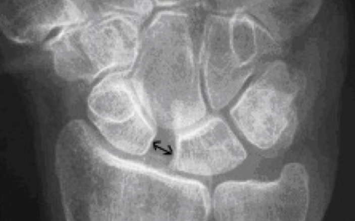
The classic finding is coined the Terry Thomas Sign. Terry was a British comedian with a gap in his teeth that nobody under sixty five really knows anymore. Today it would more appropriately be called the Michael Strahan Sign. Radiographically there is a disproportionate amount of space or “gap” noted between the scaphoid and the lunate
Normal space is 2mm or less so anything greater than 3mm is considered positive. A “ring sign” directly over the scaphoid may also be noted. The circular ring is a radiographic phenomena that occurs when the scaphoid dorsiflexes as a result of ligamentous instability. Unfortunately, it can also occur when the X-ray technician shoots the image at a slightly oblique angle, which is extremely common. Seeing the two findings together however should raise suspicion. (See figure C below)

Treatment
Scapho-lunate dissociations require surgery. It’s important for the ED physician to recognize that this injury pattern does not reflect ligamentous instability alone. With the ligament disrupted, the scaphoid subluxes dorsally. Even tiny disruptions in the normal position and alignment of the scaphoid have disastrous functional implications. Surgical reduction with repair of the scapho-lunate ligament is required to re-align these critical carpal bones. Failure to do so results in what is termed Dorsal Intercalated Segment Instability or DISI instability. This simply means that the lunate and scaphoid are out of place and the lunate shifts dorsally. Long term DISI instability is a common cause of delayed wrist fusion and advanced early onset degenerative arthritis.
Pearls
- Look for widening of the scapho-lunate space aka the “Terry Thomas” aka “Michael Strahan” sign
- Have high suspicion with a fall on an outstretched hand, dorsal wrist pain, and no obvious radiographic evidence of fracture
Luno-Triquetral Dissociations
These represent the same type of ligamentous injury affecting the luno-triquetral space. Luno-triquetral dissociations are less common because they result from forced radial deviation, which is a more unusual mechanism.
Exam
Tenderness over the luno-triquetral space along with swelling or ecchymosis should raise suspicion. The most sensitive test is palpating one finger distance distal to the ulnar head.
X-ray
Radiographically an obvious gap noted in the luno-triquetral space is indicative of injury. The lunate may displace in a volar direction.
Treatment
Luno-triquetral dissociations do not universally require surgery. Minimal deformity can be treated in a cast immobilization. Obvious volar displacement of the lunate or mal-alignment of the triquetrum requires surgical fixation. Again, when these are missed, long-term functional disability can result. The most common instability pattern is Volar Intercalated Instability or VISI instability.
Pearls
- Look for widening of the luno-triquetral space and associated pain on exam
- High suspicion when injuries result in axial load with forced radial deviation
Lunate Instability & Perilunate Dislocations
For the Emergency room physician these injuries are the most intimidating and often difficult to understand. Understanding that the lunate is the “keystone” of the wrist is critical in understanding and recognizing these injuries. The lunate is the anchor point of all carpal bones and the primary link between wrist articulation and mid-carpal movements. When things go wrong with the lunate, everything else falls apart. I outline the different injury patterns only because it helps in understanding and recognizing the injury patterns. Memorizing the names or classification is not necessary but recognizing the injury is.
Greater Arc injuries occur when fractures pass through the bones that surround the lunate. These are not subtle with fractures identified in the scaphoid, capitate, lunate and sometimes the radial styloid. This pattern represents a severe injury requiring surgery.
Lesser Arc injuries may also have associated carpal fractures (usually the scaphoid) but primarily consist of ligamentous disruptions surrounding the lunate. The sapho-lunate and luno-triquetral ligaments are disrupted along with the stabilizing soft tissue between the lunate and capitate. The two common finding in lesser arc injuries are lunate dislocations and perilunate dislocations.
Lunate dislocations are the result of lesser arc injuries and are easily recognized when the lunate is free floating volar to the carpal bones.
Perilunate dislocations are also lesser arc injuries in which the carpal bones surrounding the lunate dislocate dorsally while the lunate itself retains alignment with the radius. These may also have associated scaphoid fractures. (See figure D below)

Pearls
- Lunate dislocations occur when the lunate dislocates and all the other carpal bones maintain alignment with the distal radius.
- Perilunate dislocations involve dorsal dislocation of the carpal bones surrounding the lunate while the lunate maintains its alignment with the distal radius.
- Greater Arc injuries involve a curve like pattern of fractures passing through the bones surrounding the lunate.
The Triangular Fibro-Cartilage Complex
Physicians may look at thousands of wrist X-rays in their career without finding it unusual that there seems to be a large empty space between the distal ulna and the triquetrum. In reality, it’s not a space, and it’s not empty. It is the location of the Triangular Fibro-Cartilage Complex or TFCC. This cartilaginous structure is critical to wrist biomechanics. It extends the cup shaped arc of the carpal bones, in a manner not unlike the labrum of the shoulder, such that the carpal bones easily slide in articulation with the distal radius and ulna. It stabilizes the distal radial-ulnar joint (DRUJ) and absorbs up to twenty percent of load across the wrist.
Injury
Because the TFCC absorbs significant axial load, a fall on an outstretched hand can result in injury. Usually some degree of ulnar deviation is also part of the mechanism.
Exam
Patients present with pain on the ulnar side of the wrist immediately distal to the ulna. Like meniscal injuries they will often have clicking and pain with provocative testing, which consists of axial load and ulnar deviation.
Radiography
MRI is the standard for Diagnosis. These are not emergencies and MRI is not warranted in the ED, but clinical suspicion can help guide discharge follow up and care. When the injuries are missed patients usually go undiagnosed for years. There are radiographic findings on plain X-ray suggestive of TFCC injury. Radial shortening relative to the ulna is often noted in TFCC injuries. Up to 2.5mm of shortening can be normal but TFCC injuries usually result in 4.5mm of shortening or greater.
Treatment
Again, these are not emergencies and follow up with hand surgery is appropriate. Definitive diagnosis is made by MRI. If the ED physician fails that least recognize the possibility of this injury the patient may go years without diagnosis, Suspicion can at least help guide the patient toward appropriate treatment which is often arthroscopic wrist surgery.
Pearls
- Suspect TFCC injuries and refer to Hand Surgery when patients have ulnar side wrist pain with clicking and radiographic shortening of the radius.
Conclusion
As with most orthopedic injuries understanding functional anatomy is critical in recognizing injury patterns. Injuries to the carpal bones can be complex with life changing implications. A detailed understanding of all aspects of injury patterns and anatomy is by no means necessary for the ED physician but basic recognition of the most common injury patterns discussed here will distinguish exceptional providers and be pivotal in helping patients receive timely and appropriate treatment.
REFERENCES
- Young, Darryl, Steven Papp, and Alan Giachino. “Physical examination of the wrist.” Hand clinics 26.1 (2010): 21-36.
- Slutsky, David J., and A. Lee Osterman. Fractures and injuries of the distal radius and carpus: the cutting edge. Elsevier Health Sciences, 2009.
- Egol, Kenneth A., Kenneth J. Koval, and Joseph David Zuckerman. Handbook of fractures. Lippincott Williams & Wilkins, 2010.
- Rockwood and Green’s fractures in adults. Philadelphia: Lippincott Williams & Wilkins, 2006.
- Wolfe, Scott W., et al. Green’s Operative Hand Surgery: Expert Consult: Online and Print. Elsevier Health Sciences, 2010.
- Perron, Andrew D., et al. “Orthopedic pitfalls in the ED: lunate and perilunate injuries.” The American journal of emergency medicine 19.2 (2001): 157-162.
1 Comment
Dr. Courtney,
Nice article. FYI.
EM physicians often have to rely on their clinical skills in the ED or the field and this excellent article referenced below by Dr. Waeckerle is a great reference to include in literature on scaphoid fractures. Clinical findings result in the individual being splinted until definitive treatment is obtained. Even more amazing is that Dr. Waeckerle did this research in a private setting.
Unfortunately the medical literature is being locked up and held for ransom by the publishers so doing a literature search is becoming expensive or avoided due to the cost.
A prospective study identifying the sensitivity of radiographic findings and the efficacy of clinical findings in carpal navicular fractures
MD, FACEP Joseph F Waeckerle
Department of Emergency Medicine, Baptist Medical Center, Kansas City, Missouri, USA
http://www.annemergmed.com/article/S0196-0644(87)80563-2/pdf
SIncerely,
John Carter, MD