Intubation has unique educational challenges because of severe time restrictions and patient risk. Repetitive practice cannot be done on the same patient to separate and examine the components of the procedure in real time. As a result, most clinicians improve their skills slowly, through cumulated experience of trial and error. Intubation should involve a planned strategy to achieve first pass success, prevent hypoxemia and avoid aspiration. This article highlights two of the most common errors of direct laryngoscopy, and how to avoid them.
Intubation has unique educational challenges because of severe time restrictions and patient risk. Repetitive practice cannot be done on the same patient to separate and examine the components of the procedure in real time. As a result, most clinicians improve their skills slowly, through cumulated experience of trial and error. Intubation should involve a planned strategy to achieve first pass success, prevent hypoxemia and avoid aspiration. This article highlights two of the most common errors of direct laryngoscopy, and how to avoid them.
Error #1: Failing to appreciate the optical components of the procedure.
Direct laryngoscopy is visually challenging, with the larynx itself sighted only by the operator’s dominant pupil, at a distance of 12-18”. It is visually analogous to looking down a narrow pipe at a target the size of a quarter. The restrictions affecting direct sighting of the larynx result from the cumulative barriers of the mouth, teeth, tongue, epiglottis, and the dimensions of the laryngoscope blade. The other major variable in sighting the larynx is the operator (i.e. their specific visual acuity and light requirements). Beginning in our early forties, the near accommodation point of persons, even with a history of perfect sight, begins to change. Our near accommodation point moves 2-3 cm outward until our fifties. Reading and driving lenses are not the right distance for sighting the larynx; reading lenses are too close, and driving lenses focus too far. Continuous bifocals, or progressives, make matters even worse. As the distance to the visual target changes looking into the mouth and down to the larynx, the operator has to tilt their head and adjust the specific region of the eyeglass through which they look, otherwise the target is not in focus.
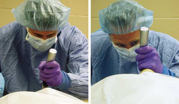
The Fix: Determine your ocular dominance and examine whether you have the right visual corrective lenses for a distance of 12 to 18 inches. To find out your ocular dominance, hold a laryngoscope with a straight blade attached and look down the barrel of the blade at a small target. Without moving your head, alternatively close one eye and then the other. When you close your dominant eye the image will move and no longer be positioned centrally at the end of the blade. When you close the non-dominant pupil the image does not move. During direct laryngoscopy we subconsciously suppress the non-dominant image because we cannot achieve stereoscopic sight (merge the right and left disparate views). About 85% of operators are right eye dominant; if you wear corrective lenses or are left-handed, there is a higher chance you will be left-eyed. [Levitan RM, et. al. Contrary to popular belief and traditional instruction, the larynx is sighted one eye at a time during direct laryngoscopy. [letter]Acad Emerg Med, 5:844-6, 1998.] [see Figure 1]. The ideal target distance is found between your left arm bent at ninety degrees and full extension. Ideal corrective lenses for laryngoscopy should not be progressives; proper lenses will not only allow you to focus at a closer distance (for those of us older than 40), but also provide slight magnification. A final consideration of laryngoscopy optics involves the illuminance of laryngoscope blades. Especially as we age, brighter laryngoscope lights make an enormous difference. Many EDs stock laryngoscope blade and handle pairs that produce inadequate light [Levitan RM, et. al. Light intensity of curved laryngoscope blades in Philadelphia emergency departments. Ann Emerg Med. 2007; 50:253-7.].

^Right and left-eyed laryngoscopists: In left-eyed laryngoscopist the head is rotated toward right (right image) aligning the left eye to the target. In right-eyed operator the head is positioned straight relative to patient (right eye aligned with target).

^Positioning a patient to achieve ear-to-sternal notch alignment:
In the top image, the head of the bed has been lifted and sheets are placed under the top of the shoulders and occiput. The face plane is parallel to the ceiling. In the lower image, a trauma stretcher is tilted to lower the foot of the bed, effectively raising the head relative to the stomach, while maintaining cervical spine precautions.
Error #2: Extending the patient’s head backward (i.e., alanto-occipital extension)
Ideal patient positioning for direct laryngoscopy involves having the patient’s face plane parallel to the ceiling, and elevating the head until the patient’s ear is horizontally aligned with the sternal notch. Imagine how patients position themselves in respiratory distress—leaning their head forward relative to the chest. We should recreate this position in a supine orientation for direct laryngoscopy [bottom]. In thin patients, a slight lift of the head of the stretcher and a towel or two will be adequate. For the morbidly obese, this may require a massive ramp under the upper back and shoulders. [Collins JS, et. al.. Laryngoscopy and morbid obesity: a comparison of the “sniff” and “ramped” positions. Obes Surg. 2004;14: 1171-5.] In patients in whom cervical spine precautions exist, the stretcher can be tilted to lower the foot of the bed, although the head cannot be raised relative to the torso [the front of the collar should always be removed and the head stabilized by an assistant].
Proper positioning is critical for finding the epiglottis. Atlanto-occipital extension pushes the epiglottis back against the posterior pharyngeal wall, making it harder to identify and harder to distract. This is a major factor in what I call “epiglottis camouflage,” where the epiglottis edge disappears against the pharyngeal mucosa. Avoiding over-extension, and having a suction tip catheter available to dab the posterior pharynx, where fluids will pool and cover the epiglottis, are both critical for “epiglottoscopy.” Also, having the head higher than the stomach is just a prudent thing in patients at risk for aspiration (i.e. full stomach). Finally, there are dramatic benefits in the efficacy of pre-oxygenation and mask ventilation with the head elevated position.
The Fix: Elevate the head to achieve ear-to-sternal notch positioning relative to progressively down the tongue and always find the epiglottis. In trauma cases with cervical spine precautions, consider tilting the foot of the bed down. Have a Yankauer suction tip ready to dab the posterior pharynx, suctioning any accumulated fluids, and be mindful of “epiglottis camouflage.”
Dr. Levitan teaches emergency medicine at Jefferson Medical College and at the Univ. of Maryland and helps run a monthly airway management course involving specially prepared cadavers: jeffline.jefferson.edu/jeffcme/Airway


