
A 23-year-old male presents after a bicycle accident. He reports falling onto his left knee, then sliding to a stop under a parked car, colliding with a grate on the street. He reports no medical problems or medications. Exam is notable for a deep laceration slightly inferior and lateral to his left patella. Bleeding is controlled; the wound base seems appreciable though somewhat limited by maceration of tissue. He has some pain with range of motion of his knee but states that his pain is mostly around the area of the laceration. Plain radiographs are negative for fracture. As you assemble laceration repair supplies, you begin to consider the possibility of knee joint involvement. How can the EP confidently rule out traumatic arthrotomy of the knee joint?
The knee is comprised of the structures that surround the bony articulations of the femur, tibia, fibula, and patella. Ligaments, cartilages, and tendons are key structures both around and outside the joint. The knee joint capsule itself can be violated by soft tissue injuries near the joint; this constitutes a surgical emergency that usually will require urgent orthopedic consultation. There is little data in the literature about what constitutes a high-risk injury aside from deeply penetrating trauma such as gunshot and stab wounds. Intra-articular gas seen on radiograph or the development of septic arthritis following a peri-articular wound are generally considered definitive signs of knee joint penetration. A high index of suspicion must be maintained for this injury.
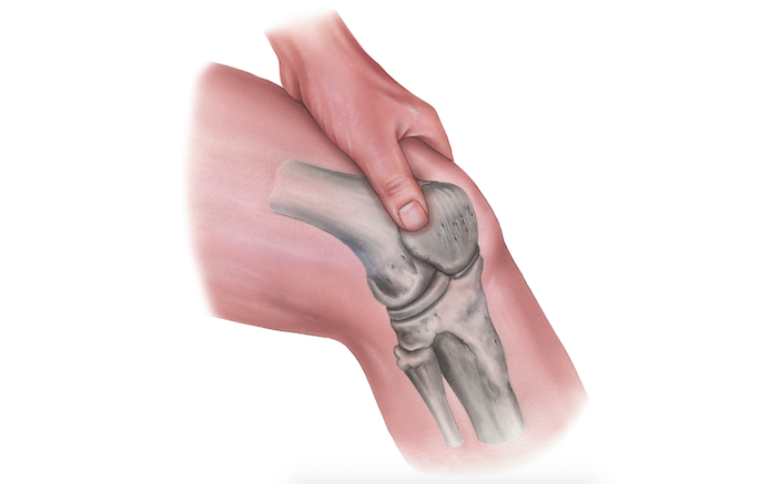
Traditionally, the saline loading test (SLT) has been a staple of investigation for possible traumatic arthrotomy. First described in the orthopedic literature in 1978, this test involves challenging the knee joint by injecting a significant amount of sterile saline into the joint space and observing for extrusion of saline from the wound(s).
Following informed consent, the following steps are taken:
- Place the knee in gentle flexion, which can be maintained with a towel roll.
- Sterilize the skin of the lower extremity from distal quadriceps to proximal calf with betadine or chlorhexidine.
- Drape the knee with sterile towels, exposing only the sterilized skin of the knee.
- Inject 2-4 mL lidocaine 1-2% (+/- epi) in a contralateral position to the injury, tracking along the planned aspiration pathway. For example, if laceration/injury inferomedial aspect of knee, inject at the superolateral aspect, tracking toward joint capsule.
- Under sterile conditions, fill a 60mL syringe with sterile normal saline (+/- 1-2 cc methylene blue).
- Attach a 20g needle to a syringe and advance carefully at the site of lidocaine injection.
- Aspirate as the needle is advanced. Confirm entry into the joint with aspiration of synovial fluid (assuming remaining synovial fluid after injury).
- After confirmation of placement, begin slowly injecting saline into joint capsule.
- As saline is injected, inspect joint for saline extrusion.
- Some authors recommend gently ranging the joint to increase visualization of extrusion of fluid.
While the procedure itself is relatively straightforward, there is debate in the orthopedic literature over how well it performs and what amount of fluid must be injected to truly rule out a small arthrotomy. In one series, a volume of 194 mL was required to achieve a 95% sensitivity for small injuries. A similar study found 95% sensitivity at a volume of 155 mL. A much smaller volume of 50 mL was less than 50% sensitive. Other studies revealed a false negative rate of 67% with volumes up to 105 mL (saline + methylene blue) and 95 mL (saline alone) and sensitivities of only 36% at volumes of 60 mL. [Metzger, Carney, Booher. J Ortho Trauma 2012]. Most orthopedic authors conclude that saline loading test alone is either insufficiently sensitive to rule out joint violation when used alone or that a significant amount of fluid must be injected to achieve adequate sensitivity. When one considers that a typical synovial fluid volume of the knee is around 7 mL [pmid 8779258], injecting 150 mL or more into that joint is a procedure that will require logistical planning and coaching of a cooperative patient.
Considering this information, the prudent EP may be left with a persistent question regarding traumatic arthrotomy and the safety of primary skin closure prior to definitive diagnostics. Given the limitations of the saline loading test, are there additional diagnostic options? There is a small body of literature indicating that computerized tomography (CT) scanning of the knee joint may have a significant role in ruling out traumatic arthrotomies. One study found that CT scanning of the joint had superior performance to saline loading test in the detection of joint injury. A cadaveric study showed that CT demonstrates greater sensitivity for even small volumes (0.1 mL) of air in the joint.
Much of the above literature reveals deficiencies of sensitivity for evaluation of traumatic arthrotomy. A positive study is clearly evident with either modality (eg SLT with extrusion of fluid, CT with free air in joint). That is to say, either study alone with a positive finding promptly concludes the diagnostic process, but either study alone with a negative finding leaves diagnostic uncertainty. The difficulty is definitively ruling out traumatic arthrotomy. There are no studies that directly compare CT to saline loading in a randomized fashion and no studies that propose a definitive algorithm combining these two modalities to exclude knee joint injury. Each diagnostic pathway provides useful information when evaluating for traumatic arthrotomy, and when available, the studies in conjunction may add to diagnostic yield.

Pro Tips: The Saline Loading Test
- 150 cc saline load into joint has high negative predictive value and 95% sensitivity in detecting small joint injuries.
- CT scan of the joint may have even greater sensitivity for small volumes of intra-articular air.
- Saline load with advanced imaging has highest sensitivity for ruling out traumatic arthrotomy.
2 Comments
Correct me if I’m wrong, but wouldn’t performing the SLT before CT cause many false positives? I feel like inserting a needle and injecting into a joint space would track air into that space and then your CT would not be properly interpretable.
More study is definitely needed to compare SLT to CT with a larger number of patients. If CT is still not sensitive enough, then maybe injecting a small amount of saline + contrast (would gastrografin be harmful if injected into a joint?) and then performing a CT yield better sensitivity?
An intraarticular injection of dilute methylene blue might provide a more easily recognized endpoint, with a smaller volume of injection.