
Check out the latest trends and best practices in the Emergency Department
Establishing peripheral intravenous (PIV) access is essential to emergency care and can often be lifesaving. However, about 35 percent of patients who present to the emergency department (ED) have difficult PIV access, particularly if traditional palpitation or landmark techniques are used, according to a recent meta-analysis.[i] With multiple failed attempts, patients suffer pain, increased risk for infection and delays in care, the investigators reported.
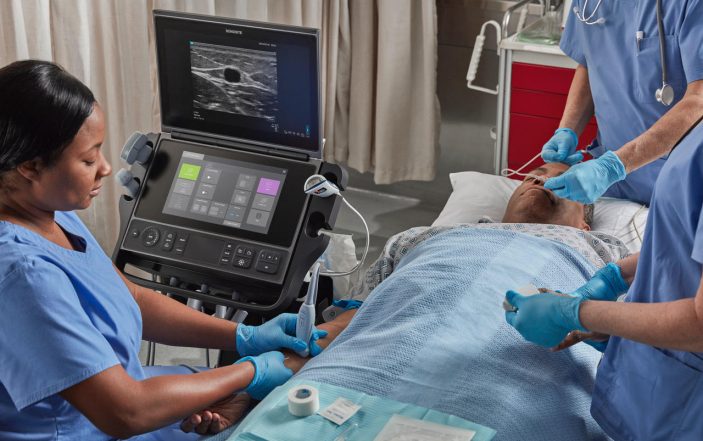
In a recent policy statement, the American College of Emergency Physicians (ACEP) recognizes the use of point-of care ultrasound (POCUS) for procedural guidance to improve the speed, safety and first-pass success rate of procedures, including PIV line placements.[ii] Subsequently, increased adoption of this technique has driven massive changes, quality improvements and potential cost savings in emergency medicine (EM) practice.
This review offers an overview of best practices and current trends in the use of ultrasound-guided PIV (USPIV), drawing on recent literature and the author’s experiences as the division director of emergency ultrasound at Vanderbilt University Medical Center. The value of having an interprofessional team of EM providers trained in the technique is also highlighted.
A Safer Alternative to High-Risk Central Lines
Increased use of USPIV is helping EM providers address one of the biggest—and most dangerous—bottlenecks in emergency care: difficulty in obtaining PIV access. In recent years, many EDs across the US, including ours, have seen a large rise in patients who present with challenging vasculature due to obesity, IV drug abuse, chronic illness and other factors. The result can be an “access molasses,” delays in essential treatment for seriously ill patients who need it the most. Failure rates of emergent PIV cannulation of 10 to 40 percent have been reported in the literature if traditional techniques are used. [iii]

In this scenario, it used to be common for clinicians to turn to central venous catheters (CVCs) as an alternative. However, CVCs can have significant dangers, including pneumothorax, hemothorax, thrombosis, hemorrhage, air embolism and central line-associated bacterial infections (CLASBIs). An estimated 250,000 CLASBIs occur in U.S. hospitals, with an associated estimated cost of up to $94,000 per case, according to a meta-analysis by the Agency for Healthcare Research and Quality.[iv]
The Centers for Medicare & Medicaid Services have given hospitals a powerful incentive to reduce CLASBIs by imposing financial penalties on facilities with the highest rates of these and other hospital-acquired conditions. A large body of research has shown that USPIV can dramatically improve patient safety, and cut costs, by helping those with difficult PIV access avoid unnecessary CVCs. For example, the meta-analysis cited above found that in such patients, PIV cannulation was four times more likely to be successful with ultrasound guidance than without it.
Safely Accelerating Lifesaving Emergency Care

Many investigations have demonstrated important benefits of USPIV in the ED setting, where faster treatment helps improve outcomes. Until a line has been successfully placed, many common therapies cannot be initiated, including the delivery of lifesaving medications, fluids and blood products, volume resuscitation, hemodynamic monitoring, and hemodialysis. Similarly, PIV access is vital for diagnosis, allowing blood to be drawn for laboratory analysis and contrast agents administered for imaging studies; and is a prerequisite for transferring patients to other departments for further treatment, such as surgery, cardiac catheterization, or critical care.
A study of 100 ED patients with difficult vascular access reported a 97% success rate with ultrasound guidance, compared to only 33% when traditional landmark approaches were used. USPIV was also found to achieve faster vascular access (13 minutes versus 30 minutes without ultrasound) with fewer punctures (an average of 1.7 versus 3.7 with landmark methods). Another comparative study of difficult-access ED patients found that ultrasound guidance eliminated the need for CVCs in 85% of cases.[v] The patients were tracked for seven days, with zero complications in the ultrasound group and a 6.7% rate of CLASBI in the landmark group.
An Innovative Strategy for the Management of Septic Shock
The growing trend to reduce the use of CVCs whenever possible—and thus avoid their high costs and many dangerous complications—have sparked scientific inquiry into new roles for PIV catheters. For example, vasopressor medications for the management of septic shock have traditionally been administered via CVC due to concerns about the risk for tissue injury and necrosis if extravasated. However, the need for these devices has been questioned, leading to a 2020 systematic review analyzing the potential for delivering vasopressors peripherally.
The review revealed that it may be safe and feasible to administer vasopressors peripherally, if the treatment is of short duration.[vi] The researchers analyzed seven studies that included 1,382 patients, none of whom developed tissue ischemia or limb necrosis during a mean duration of 22 hours of infusion. Extravasation occurred in 3.4% of patients, all of whom were successfully treated conservatively or with vasodilatory medications.
A Visual GPS to Locate the Safest Peripheral Catheter Sites
Here is how real-time ultrasound visualization helps solve the problem of vascular molasses: It allows EM providers to map the patient’s veins, access their patency, and quickly identify the best target for efficient cannulation. In addition, ultrasound guidance allows clinicians to evaluate vein size, with a vessel width of 0.4 cm being associated with 60 percent higher cannulation success rate, as compared to veins with a smaller width, Witting and colleagues reported.[vii] Success rates were also highest in vessels of moderate depth (0.3 to 1.5 cm).
Along with facilitating faster PIV cannulation of superficial veins that are common targets for palpation methods (such as the forearm, antebrachial, and median cubital veins), POCUS also permits visualization and guidance for deeper veins that cannot be located with palpation. For example, if no forearm vein is adequate for cannulation, the ultrasound probe can be advanced to the patient’s upper arm to visualize the cephalic and basilic veins, both of which represent ideal targets for USPIV. [viii]
In visualizing veins of the upper arm for potential PIV placement, the cephalic vein often offers a readily accessible target due to its superficial course along the anterior area of the bicep. Should this vein not be adequate, the basilic vein, which frequently widens as it is traced proximally, also has important advantages for USPIV. Too deep to be found with palpitation, the basilic vein offers a very safe forearm target because it is not associated with an artery, thus reducing risk for accidental injury to adjacent structures as the catheter is placed.
Leveraging a Team Approach to Improve Patient Outcomes and Satisfaction
To help ensure that ED patients treated at the center where the author practices have around-the-clock access to many proven benefits that USPIV offers, all 46 of our emergency physicians, as well as 20 medics and 8 registered nurses, are trained in the technique. Having an interprofessional team, which could also include nurse practitioners and physician assistants, significantly enhances rapid, cost-efficient workflow. Along with procedural ultrasound’s well-established advantages for accelerating lifesaving emergency care, and reducing medical mistakes, it has also been shown to increase patient satisfaction.
As the value of USPIV become increasingly widely recognized by both EM providers and the public, our ED has seen a massive increase in the number of patients who ask for it upon arrival. Often, they will say, “Doc, I’m a difficult stick” and some chronically ill patients will even tell us which blood vessels worked best the last time they were treated at our center. And when patients turn to us for help in an emergency, ultrasound is at the bedside, ready to facilitate the leading-edge care our patients need—and deserve.
References
[i] Stolz LA, Stolz U et al, Ultrasound-guided peripheral venous access: a meta-analysis and systematic review. J Vasc Access, 2015; 16 (4):321-326.
[ii] Emergency Ultrasound Imaging Criteria Compendium. Ann Emerg Med, Vol. 68, Issue 1, e11-48. Available at https://www.annemergmed.com/article/S0196-0644(16)30096-8/fulltext.
[iii] Leidel BA et al. Is the intraosseous access route fast and efficacious compared to conventional central venous catheterization in adult patients under resuscitation in the emergency department? A prospective observational pilot study. Patient Saf Surg. 2009 Oct 8;3(1):24.
[iv] Agency for Healthcare Research and Quality. Estimating the Additional Hospital Inpatient Cost and Mortality Associated With Selected Hospital-Acquired Conditions. Available at https://www.ahrq.gov/hai/pfp/haccost2017-results.html
[v] Au AK, Rotte MJ, Grzybowski RJ, Ku BS, Fields JM. Decrease in central venous catheter placement due to use of ultrasound guidance for peripheral intravenous catheters. Am J Emerg Med. 2012 Nov;30(9):1950-4.
[vi] Tian DH, Smyth C, Keijzers G, Macdonald SP, Peake S, Udy A, Delaney A. Safety of peripheral administration of vasopressor medications: A systematic review. Emerg Med Australas. 2020 Apr;32(2):220-227.
[vii] Witting MD, Schenkel SM et al. Effects of vein width and depth on ultrasound-guided peripheral intravenous success rates. J Emerg Med. 2010 Jul;39(1):70-5.
[viii] Presley B and Isenberg JD. Ultrasound Guided Intravenous Access. [Updated 2021 Jul 31]. In: StatPearls [Internet]. Treasure Island (FL): StatPearls Publishing; 2022 Jan. Available at https://www.ncbi.nlm.nih.gov/books/NBK525988/
1 Comment
I am an older ED physician provider. I do not have ultrasound skills. How does one become proficient in ultrasound at a later age?
I am ABEM certified in EM .