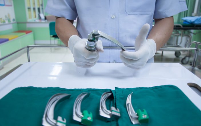
Is direct laryngoscopy a dying skill?
“I will unequivocally state that it is wrong for people to practice direct laryngoscopy in 2012.”
— Ron Walls on EMRAP September 2012.
Those were awfully harsh words from the man who literally wrote the book on emergency airway management. This statement understandably caused a considerable amount of controversy at the time, and almost a decade later, direct laryngoscopy is still practiced in emergency departments, though its use is on the decline.[1]
Is it just a matter of time before direct laryngoscopy is considered as outdated and crude of a device as Ron Walls claimed in his infamous appearance on EMRAP in 2012? Does video laryngoscopy have the potential to become the be-all and end-all of emergency airway management? In order to settle the debate of video vs. direct laryngoscopy, let’s consider the following three questions:
- Who wins in the battle for first pass success?
- Can emergency physicians master both DL and VL?
- Is VL a sufficient replacement for DL?
Before jumping in, it is crucial to clarify some important terminology. A “standard geometry” or Macintosh video laryngoscope is essentially a Macintosh direct laryngoscope with a camera attached. It is often considered synonymous with the popular Karl Storz C-MAC video laryngoscope system, and can be used to obtain a direct view of the glottis or indirect view with the video camera.
Hyperangulated video laryngoscopes, on the other hand, feature a blade with a hyper-acute angle. The Glidescope system is colloquially synonymous with said laryngoscope though several other brands exist as well. Its use requires the view obtained by the video camera, as the hyperangulation of the blade makes direct visualization of the airway impossible.
Today, both the C-MAC and Glidescope systems feature standard geometry and hyperangulated blades. With that clarification out of the way, let us consider the first question.
Who Wins the Battle for First Pass Success?
In order to answer this, let’s first take a look at Ron Walls’ own data. [1] The National Emergency Airway Registry is a cohort of 18 EDs in the United States, Canada and Australia. Its data was published in Annals of Emergency Medicine in 2015 and provides some important insight into the trends of emergency airway management over time. The data show that video laryngoscopy use began to increase after 2006, with an initial increase in first pass success (FPS).
However, FPS plateaus after 2007, despite a dramatic and sustained increase in the use of video laryngoscopy. If VL was clearly superior to DL, why has its increased use not led to a further increase in first pass success in this cohort?
Let’s turn to the “highest” forms of evidence — the systematic review and meta analysis. A 2016 Cochrane Review of 64 randomized controlled trials of 7,044 patients found that video laryngoscopy decreased failed intubation compared to direct laryngoscopy. [2]
However, it found no difference in first pass success, number of attempts, hypoxia and mortality. Additionally, while a 2018 meta-analysis of five randomized controlled trials with 1,250 patients showed that VL decreased esophageal intubation, it also found no difference in first pass success as well as overall success, time to intubation, and survival to discharge. [3]
If video laryngoscopy does indeed lead to decreased failed and esophageal intubation, it reasons that these devices are a useful “bail-out” when direct laryngoscopy fails. However, if they do not lead to increased first pass success, then why would we be obligated to use them preferentially for our first attempt?
Can we be competent at both?
The Accreditation Council for Graduate Medical Education (ACGME) requires that emergency medicine residents perform at least 35 intubations in order to graduate. Where does the ACGME get this number? Apparently not from the evidence.
A systematic review performed in 2015 said that at least 50 intubations are needed to achieve a 90% success rate within two attempts in elective circumstances. [4] A study by Bernhard et al. showed that first pass success steadily increased from 67% within the first 25 intubations to 83% after 200 intubations. [5] Importantly, the vast majority of this evidence comes from studies of direct laryngoscopy.
These numbers come with important caveats. We care about first pass success, not success within two attempts. We also do not work under elective circumstances. Emergency airway management is complicated by factors including, but not limited to uncertain NPO status, airway contamination with blood and emesis, hypoxia and hemodynamic instability. We can conclude that the true number for competence is likely much higher than 50 when it comes to emergency intubation.
So how does the experience of EM residents compare? A 1999 national survey showed that EM residents performed an average of 75 intubations during their three- or four-year residency. [6] A more recent single-center study found that residents performed an average of 28.91 intubations per year during their three-year residency. [7] Therefore, in order to master direct laryngoscopy, EM trainees must prioritize its use during residency.
What about video laryngoscopy? Standard geometry VL overlaps in technique with direct laryngoscopy because it uses the same blade. As the video camera is known to provide a higher grade Cormack-Lehane view, [8] it therefore reasons that those competent with DL will be able to perform standard geometry VL.
That leaves us with hyperangulated video laryngoscopy. Because of its blade geometry, hyperangulated VL requires a separate technique than that with a Macintosh blade. Therefore, it reasons that the more trainees use these devices, the less they progress along the steep, but prolonged learning curve of gaining competence with direct laryngoscopy.
The good news is you can learn to use hyperangulated VL on a mannequin. One study looked at 20 trainees who had never performed an intubation. They were trained with a Macintosh direct laryngoscope and hyperangulated GlideScope on airway models until they could perform each technique successfully three times in a row. They then attempted 10 intubations on real patients; five with DL and five with a GlideScope. Cumulatively, the GlideScope was successful 93% of the time compared to 51% with DL. [9]
I believe hyperangulated video laryngoscopy is easily learned on mannequins because it is a non-displacement laryngoscope. In order to perform the technique, alignment of the oral, pharyngeal and laryngeal axes is not required to view the glottis with the camera, unlike with direct laryngoscopy. This minimizes the discrepancy between intubating a mannequin and a real patient.
No tongue sweep is required, nor sniffing position, lifting up and away to displace the lingual, oropharyngeal and submandibular tissues. The patient is positioned with a neutral spine, and the blade is inserted along the midline of the tongue until the airway is revealed with the camera. Plastic mannequin “tissue” does not behave the way real human tissue does, and that’s why direct laryngoscopy (a displacement laryngoscope) is harder to learn on airway models and requires training with real patients in order to attain mastery.
Where does VL fall short?
If video laryngoscopy is the way of the future, is it a sufficient replacement for DL? I would argue no. If the camera malfunctions due to technological failure or contamination with blood or emesis, you can at least try to rescue your attempt with direct visualization if you use a standard geometry blade, although you must be competent at direct visualization to use this as your backup. If you use a hyperangulated blade, the device is useless if the camera view is lost.
While I was unable to find literature on how often this occurs, in my experience it is too much. During my first six months of my residency, the camera shut off in the middle of two separate intubation attempts with a Glidescope. The results were not pretty. And in my emergency department, we only have one video laryngoscope, leaving us with direct laryngoscopy as our only backup when VL inevitably fails.
Also, because a hyperangulated does not align the axes of the airway, any foreign body removal proves to be much more difficult. In a cadaver study comparing the Macintosh direct laryngoscope to the GlideScope, foreign body removal was 100% successful on the first pass with DL, while the GlideScope was only 78.6% successful. More strikingly, direct laryngoscopy allowed removal of the foreign body in a median of 16 seconds, compared to 84 seconds with video laryngoscopy. [10]
In Conclusion
The debate between DL and VL is easily over-simplified because standard geometry video laryngoscopy offers the best of both worlds. Using these devices to obtain direct visualization allows trainees to practice and maintain the skill of direct laryngoscopy. If an adequate view cannot be obtained, we can simply turn towards the video screen which will afford us a better view. In this way, our first two attempts are built into one, thereby maximizing our chances of success.
While video laryngoscopy may decrease esophageal intubation, I would argue that a quick verification of correct placement on the video screen after tube delivery under direct visualization would eliminate this difference.
We emergency physicians consider ourselves masters of resuscitation. What is the essence of resuscitation? The ABC’s: Airway, Breathing and Circulation. If we want to own resuscitation, we have to own the airway. If that is the case, why would we ever remove from our skill set the technique that has proven itself to be highly reliable and successful ever since its inception in 1943? Is DL dead? No, and emergency physicians need to continue to learn, practice and embrace this essential skill.
References:
- Brown 3rd CA, Bair AE, Pallin DJ, Walls RM, NI Investigators. Techniques, success, and adverse events of emergency department adult intubations. Ann Emerg Med 2015;65:363–70, e1.
- Lewis SR, Butler AR, Parker J, Cook TM, Schofeld-Robinson OJ, Smith AF (2017) Videolaryngoscopy versus direct laryngoscopy for adult patients requiring tracheal intubation: a Cochrane systematic review. Br J Anaesth 119:369–383
- Bhattacharjee S, Maitra S, Baidya DK. A comparison between video laryngoscopy and direct laryngoscopy for endotracheal intubation in the emergency department: A meta-analysis of randomized controlled trials. J Clin Anesth. 2018;47:21-26. doi:10.1016/j.jclinane.2018.03.006
- Buis ML, Maissan IM, Hoeks SE, Klimek M, Stolker RJ. Defining the learning curve for endotracheal intubation using direct laryngoscopy: A systematic review. Resuscitation. 2016;99:63-71. doi:10.1016/j.resuscitation.2015.11.005
- Bernhard M, Mohr S, Weigand MA, Martin E, Walther A. Developing the skill of endotracheal intubation: implication for emergency medicine. Acta Anaesthesiol Scand. 2012;56(2):164-171. doi:10.1111/j.1399-6576.2011.02547.x
- Hayden SR, Panacek EA. Procedural competency in emergency medicine: the current range of resident experience. Acad Emerg Med. 1999;6(7):728–35.
- Bucher JT, Bryczkowski C, Wei G, et al. Procedure rates performed by emergency medicine residents: a retrospective review. Int J Emerg Med. 2018;11(1):7. Published 2018 Feb 14. doi:10.1186/s12245-018-0167-x
- Brown 3rd CA, Bair AE, Pallin DJ, Laurin EG, Walls RM, National Emergency Airway Registry Investigators. Improved glottic exposure with the Video Macintosh Laryngoscope in adult emergency department tracheal intubations. Ann Emerg Med 2010;56:83–8.
- Nouruzi-Sedeh P, Schumann M, Groeben H. Laryngoscopy via Macintosh blade versus GlideScope: success rate and time for endotracheal intubation in untrained medical personnel. Anesthesiology. 2009;110(1):32-37. doi:10.1097/ALN.0b013e318190b6a7
- Je SM, Kim MJ, Chung SP, Chung HS. Comparison of GlideScope(®) versus Macintosh laryngoscope for the removal of a hypopharyngeal foreign body: a randomized cross-over cadaver study. Resuscitation. 2012;83(10):1277-1280. doi:10.1016/j.resuscitation.2012.02.032


