
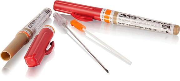
Figure above: A 14g Chest Decompression Needle kit from North American Rescue.
You’re working an overnight shift in the community hospital emergency department. A 42-year-old male farmer fell 20 feet from the barn loft and presents holding his left wrist. He is tachycardic, tachypneic, slightly hypotensive, and appears to be in moderate distress. On your primary survey, you note decreased breath sounds on the left. On your extended FAST exam you detect no lung sliding on the left. You reach for the 14 gauge angiocatheter as you visualize the 2nd intercostal space in the midclavicular line, when your colleague, an EP with recent frontline experience from the war in Afghanistan, gives you a better idea.
Medicine from the front

For more than a decade, the US military has focused on the major preventable causes of death in combat such as compressible hemorrhage, traumatic airway obstruction and tension pneumothorax. Military research has revolutionized battlefield medical care, and many military techniques can translate to the management of civilian trauma care.
Tension pneumothorax is rare in civilian trauma care settings, but is one of the leading causes of preventable death on the modern battlefield[1,2]. Without treatment, tension pneumothorax rapidly progresses to shock and death, and the first emergency treatment of choice is needle decompression.
Autopsy studies of chest wall thickness in military service members demonstrate that at least a 3.25 inch, 14-16 gauge angiocath is required to penetrate the chest wall and pleural cavity in most military casualties[3,4,5]. This information is relevant to the civilian EMS and ED patient populations, since treatment must accommodate patients with all types of body habitus.

The standard civilian recommendation for needle decompression is needle placement into the affected side of the chest, at the second intercostal space in the midclavicular line, just above the rib to avoid the intercostal artery[6]. However, several military studies support an alternative approach in the fourth or fifth intercostal space at the anterior axillary line [3,7,8]. This placement recommendation is based on the relatively thinner chest wall at the anterior axillary line and the decreased chance of injuring vital organs in the lateral chest compared to the central chest [8]. (Figs 2 and 3)
A recent military study of postmortem CT and autopsy results demonstrated a lower treatment failure rate in the lateral approach compared to the anterior approach[3]. Though the study was small, it certainly provided a compelling perspective. If the lateral approach is potentially safer and more effective for the combat medic, it may also be useful in the ED.

Figure 2: Traditional decompression into the second intercostal space at the midclavicular line. Needle has entered the lung itself.


Figure 3: Decompression at the 4th intercostal space, anterior axillary line.
Case Conclusion
In the resuscitation room, your colleague assists as you identify the fourth intercostal space at the anterior axillary line, and you insert a 3.25 inch 14 gauge angiocatheter to decompress the patient’s chest. The patient’s clinical condition improves markedly as you complete your initial evaluation and progress through your secondary survey. The successful needle decompression provides the time to perform the tube thoracostomy. Later, as the patient is packaged for admission to the surgical service, you reflect about your colleague’s unique experiences and realize that emergency medicine is indeed a nexus where many paths meet, including battlefield experience gained in Iraq and Afghanistan.
Step By Step
1. Place the patient in either lateral recumbent position with the affected side up, or supine, with the head of the bed up 40-45 degrees.
2. Identify the fourth or fifth intercostal space in the anterior axillary line.
3. Prep the area.
4. Insert the 14 or 16 gauge angiocatheter with needle placed just above the rib, perpendicular to the skin. As you traverse the pleura, you may hear the distinctive rush of air from the decompressed tension pneumothorax. Some EPs will attached a 10cc syringe partially filled with saline or water to the end of their angiocath/needle set. This allows them to visualize the “rush of air” which may otherwise not be heard in a noisy trauma bay.
5. Remove the needle and leave the catheter in place, securing it to prevent dislodgment.
6. Re-evaluate the patient to ensure a positive clinical effect and continue
to monitor the patient closely as you complete the evaluation and resuscitation.
7. Now that the tension pneumothorax has been converted to a simple pneumothorax, you can perform a tube thoracostomy.
REFERENCES
1. McPherson JJ, Feigin DS, Bellamy RF. Prevalence of tension pneumothorax in fatally wounded combat casualties. J Trauma. 2006;60:573-8.
2. Holcomb JB, McMullen NR, Pearse LA, et al. Causes of death in Special Operations Forces in the Global War on Terror. Ann Surg. 2007;245:986-91.
3. Harcke HT, Mabry RL, Maxuchowski EL. Needle Thoracentesis Decompression: Observations from postmortem computed tomography and autopsy. J Spec Ops Med. 2013;13:53-8.
4. Givens ML, Ayotte K, Manifold C. Needle thoracostomy: implications of computed tomography of chest wall thickness. Acad Emerg Med. 2004;11:211-3.
5. Harcke, HT, Pearse LA, Levy AD, Getz JM, Robinson SR. Chest wall thickness in military personnel: implications for needle thoracentesis in tension pneumothorax Milit Med. 2007;172:1260-3.
6. Kirsch, TD Sax, J. “Tube thoracostomy,” in Roberts and Hedges’ Clinical Procedures in Emergency Medicine 6th Ed., 2013; pp. 191-211.
7. United States Army Institute of Surgical Research. Tactical Combat Casualty Care Guidelines, Published 17 September 2012. https://www.jsomonline.org/TCCC/TCCCGuidelines20120917.pdf
8. Inaba K, Ives C, McClure K, et al. Radiologic evaluation of alternative sites for needle decompression of tension pneumothorax. Arch Surg, 2012;147(9):813-8.
7 Comments
What’s so new or unique about this?? Most civilian docs who deal with such have used these techniques for as long as I can recall (since 1955).
Reasonable question, Jon.
If it has been around since 1955, how come ATLS, Roberts and Hedges, and virtually all other training for needle decompression has reinforced the 2nd intercostal space, mid-clavicular line?
The fact is that this approach is not mainstream and bears mentioning.
If most civilians were already doing this today, the editorial staff would not have flagged this topic as unique or interesting.
This is just two military EP’s sharing some professional experience…why the negative vibe?
Its new in that traditional residency teaching is that needle decompression be done at 2nd IC space, MC line whereas tube thoracost. is at 4th IC space, AA line. This technique advocates needle decompression laterally as a possibly better location. Makes sense to me and NOT the way I was taught.
We have for years used a Turkel needle for this purpose. It has several advantages:
1. it is longer, 2 it doesn’t kink after insertion, 3. it has a clear indication you are in the chest (color red to green), and 4 has holes on the side as well as the tip. Note I have no financial interest in this device and there may be other similar devices I am not aware of
Jon, you’re correct in that the technique hasn’t changed. However I think the point is more about needle selection. Most EDs still don’t stock the 3.25 inch needles necessary to effectively decompress a PTX.
We actually stock 3.25 inch 10g angiocaths just for this reason – I haven’t had a PTX I haven’t been able to hit yet with those things.
I agree.Working with a peds trauma transport team, A medic-RN insisted on the classic mid-clavicular approach to a critical 4 yr-old,what came back was not air, but lymph-appearing fluid. Much safer with the anterior axillary approach.
This article misses out various key points. The procedure is fairly simple but the diagnosis clinically is not easy. The respiratory rate is rapid and the patient is distressed if conscious. They do not want to co-operate with examination because they are hypoxic. They are probably verbalising and pulling at their oxygen mask. The hyper resonant percussion note is hard to detect and the affected side is not silent as sounds are transmitted across the chest. The trachea often does not deviate, and because they won’t cooperate eliciting reduced movement on the affected side is difficult. If the person is fat its even more difficult. Go with your instincts.
If the person is unconscious respiratory rate can be depressed due to a head injury, not elevated. Ventilation of course increases the risk of a tension. Its usually a nurse or technician ventilating them. A key sign of trouble is when the person ventilating starts messing around with their mask grip, putting an OP in, trying suction etc. They know they aren’t making the chest rise, but they assume its their fault, not that something has gone wrong with the patient. If you see them do this examine the patient NOW.
They say the needle should go over the rib below and not below the rib above to avoid the neuromuscular bundle. Great in theory but with a distressed patient and a respiratory rate of 40+ that is more wishful thinking than reality. If you miss the intercostal space and hit rib when you are trying to needle the chest, go upwards, not down. Use the biggest needle you can! The Thoraquik and the ARS are the only two devices the size you need.
Don’t expect the patient to rapidly improve. It’s a myth! Try blowing throw one of those things! It takes minutes for the pressure to dissipate, not seconds. I’ve put a chest drain in three minutes later and released residual pressure.
If you didn’t get a hiss of air, and you probably won’t hear one in the prehospital environment, reassess, and if you still think its a tension make a finger sized thoracostomy. Mea culpa! I learned the hard way and two patients died as a result. Chest walls can be VERY thick.
Finally, don’t trust the device. Moving patients is a pretty good way of pulling the cannula end out of the chest cavity, and then they quietly deteriorate on you again.
If you read this, you may avoid some of the mistakes I made in 25 years of prehospital emergency care. Good luck!