In Leviticus 13:43-44, the Bible references a man with a reddish-white swollen infection on his bald head, calling him a “leprous man” and “unclean.” Thousands of years later, the CDC is warning of “rising evidence” of leprosy in the United States and has suggested that leprosy may now have become an “endemic disease process” in central Florida.
Mycobacterium leprae was discovered in 1873 by the Norwegian physician Gerhard Hansen and was the first bacterium to be identified as a cause of disease in humans. Leprosy, also called Hansen disease, is a chronic infectious disease caused by Mycobacterium leprae. Ninety-five percent of all new leprosy cases are reported from 14 countries.
In 2019, the highest incidence was seen in India, Brazil, and Indonesia. The incidence of leprosy in the United States has persistently increased since 2000. Almost one-fifth of reported leprosy cases in the United States come from central Florida. There is some evidence suggesting zoonotic transmission of leprosy since many unrelated human leprosy cases had a unique strain of M. leprae carried by armadillos in the same region.
The CDC has also stated that the increase in migration to the United States may be linked to an increased incidence of leprosy in nonendemic areas, but that the incidence of new leprosy diagnoses in foreign-born individuals is decreasing while the incidence of new leprosy diagnoses in Florida is significantly increasing.
Mycobacterium leprae is extremely slow-growing, dividing approximately every 12 days. Therefore, the incubation period for leprosy is estimated to be between 9 months and 20 years, averaging about 4 years. Because of this long incubation period, the mode of transmission is difficult to determine. Leprosy is most likely transmitted by aerosol or by direct contact with ulcerated nodules. It may also be transmitted by contact with infected nine-banded armadillos or red squirrels which both serve as natural reservoirs for M. leprae. Leprosy is not transmitted by casual contact. Asymptomatic latent leprosy infections are common in endemic areas, may persist for many years, and may resolve at any time. The manifest disease occurs in only 5–10 % of infected individuals since approximately 95% of the population is naturally immune to the disease.
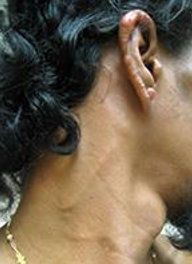
Leprosy initially presents as an “indeterminate” form characterized by the appearance of one or more pale patches on the face, ears, or limbs. These patches are non-specific and are often mistaken for other skin conditions such as hives, fungal infections, skin flaking, or loss of skin pigmentation.

Patient with indeterminate leprosy. Note visibly enlarged greater auricular and cervical cutaneous nerves on the patient’s neck. Also, note multiple reddish lesions on the patient’s ear. Picture from CDC report.
Even if leprosy is suspected, a biopsy of a lesion usually does not yield specific results, and the leprosy-causing bacterium, Mycobacterium leprae, cannot be grown in a lab setting. PCR testing may be used for diagnosis, but its accuracy in the early stages of the disease is not reliable. Special staining techniques can sometimes reveal the presence of acid-fast bacteria within certain cells or nerves in the skin, which suggests leprosy.
Because M. leprae has an affinity for Schwann cells in nerve sheaths, two of the cardinal signs of leprosy involve nerve function: 1) loss of sensation in a hypopigmented or reddish skin patch or 2) a palpably thickened or enlarged peripheral nerve with loss of sensation or weakness of the muscles supplied by that nerve. Both of these findings may be early clues to a leprosy diagnosis, although neither reliably occurs early in the course of the disease. Asymmetric neurologic abnormalities may also be present even in the absence of skin lesions.
If indeterminate leprosy progresses, it is divided into three main subtypes: Paucibacillary (tuberculoid) leprosy which is associated with good host immunity, multibacillary (lepromatous) leprosy which is associated with poor host immunity, and an intermediate form which may show varying degrees of clinical features from both tuberculoid and lepromatous leprosy.
Tuberculoid leprosy presents as solitary papules which may coalesce into erythematous plaques having raised borders and an annular appearance. The lesions usually occur on the extremities and may exhibit decreased sensitivity to pain and temperature. Intraneural granuloma formation may cause pressure-induced atrophy with loss of distal neural function. Nerve granuloma may also undergo necrosis and ulceration/abscess formation.
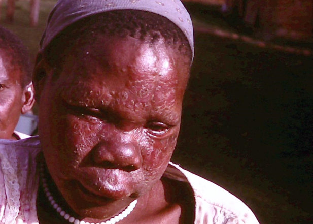
Lepromatous leprosy is a condition that affects individuals with weakened T-cell immunity. It is characterized by the appearance of multiple red-brown nodules on the skin and mucous membranes. The face, ears, and extremities are particularly vulnerable to these infiltrates.
Over time, the facial nodules can lead to “leonine facies,” marked by swollen cheeks and the loss of eyebrows and eyelashes. The presence of a high bacterial load can impact various bodily functions, including vision, kidney and liver function, and joint mobility. The disease also damages peripheral nerves, resulting in visibly enlarged nerves and peripheral neuropathy. In some cases, bone loss may occur, leading to severe deformities or even the need for amputation.
The multidrug treatment regimen recommended by the WHO includes dapsone, rifampin, and clofazimine for 12 months. Monotherapy with dapsone is obsolete due to resistance. Dapsone has a bacteriostatic effect on M. leprae, rifampin has bactericidal effects on M. leprae, and clofazimine is predominantly anti-inflammatory but weakly bactericidal.
Active surveillance, contact tracing, and research on transmission routes are important for identifying sources and reducing the spread of leprosy. The WHO also recommends the administration of a single dose of rifampin to social contacts of patients diagnosed with leprosy as preventive chemotherapy.
The CDC now suggests that travel to central Florida, even in the absence of other risk factors, should prompt consideration of leprosy in the appropriate clinical context.
Summary
- Indeterminate leprosy is difficult to diagnose
- That non-resolving rash from your patient’s pet armadillo may not be urticaria
- Cardinal signs of leprosy include:
- Loss of sensation in a pale or reddish skin patch or
- A thickened/enlarged peripheral nerve with atrophy or neuropathy distal to the site
- Leonine facies are suggestive of the multibacillary form of leprosy. Remember leonine lepromatous leprosy
- Although rarely diagnosed, leprosy may now be endemic in central Florida
References
Bhukhan A, Dunn C, Nathoo R. Case Report of Leprosy in Central Florida. Emerging Infectious Diseases. 2023;29(8):1698-1700. DOI:10.3201/eid2908.220367.
Fischer M. Leprosy – an overview of clinical features, diagnosis, and treatment. Journal of the German Society of Dermatology, 2017. DOI:10.1111/ddg.13301
World Health Organization. Leprosy, 2023. https://www.who.int/news-room/fact-sheets/detail/leprosy
Centers for Disease Control and Prevention. Hansen’s Disease (Leprosy) 2023. https://www.cdc.gov/leprosy/health-care-workers/clinical-diseases.html
Photo Credit:
[Main] Patient with lepromatous leprosy and “leonine facies”. Picture by Rosemary A Jones – Own work, CC BY-SA 4.0, https://commons.wikimedia.org/w/index.php?curid=50645849


