How to recognize and appropriately manage a dangerous snakebite
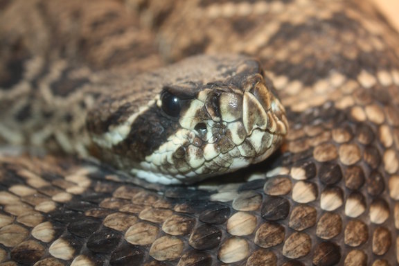
Earlier this year, during a subzero blizzard with “white-out” conditions, a 48 year old male patient presented to an urban hospital emergency department (ED) with a complaint of being bitten by an Eastern diamondback rattlesnake, Crotalus adamanteus 30 minutes prior to arrival (Figure 1). The patient had been performing in a traveling reptile exhibit, when he “tripped and fell” while handling the snake, resulting in a bite to his right hand.
As per the reptile exhibit snakebite protocol, the patient was given “a shot of epinephrine” and the 911 System was activated. Soon after, advanced life support paramedics rapidly transported the patient to the closest hospital. Upon arrival to the ED, the patient reported hand pain and swelling but denied systemic symptoms.
The patient had a past medical history significant for coronary heart disease and hypercholesterolemia. He was taking one 81mg aspirin daily, but was not on any other anticoagulant therapy. The patient denied recent alcohol consumption or illicit drug use.

Figure 2: Rattlesnake bite
Pertinent Physical Findings
Vital signs:
- Temperature = 36.5 C
- General appearance: The patient was alert, oriented X3, anxious and in moderate distress
- Extremities: Right hand with moderate edema compared to left hand, erythema with two dorsal puncture wounds and abrasions (Figure 2). Brisk capillary refills were noted with strong distal pulses. Lymphangitis was observed tracking from volar aspect of the wrist to the elbow.
- Neurological: Decreased hand grip strength with hyperesthesia through-out the involved hand
- Abdomen: Tenderness to palpation in the left lower quadrant. It was discovered at this time the patient had been bitten a second time on the abdominal wall, with superficial ecchymosis and two small puncture wounds. (Figure 3)
Laboratory Data
- Complete blood count: WBC: 9.3 103/mcL; Hgb: 9.9 g/dL Platelets: 481 mc/L
- Sodium:136 meq/L; Potassium 3.8 meq/L; Chloride: 104 meq/L; bicarbonate: 21 meq/L
- PTT: 31.3sec; PT: 12.3sec; INR 1.1; Fibrinogen: 379mg/dL (normal range 150-450mg/dL); Fibrin Split Products (FSPs): Negative; CPK: 16 U/L
- ECG: Sinus tachycardia 104 beats/minute; no ST segment elevations or depressions, no Q waves, no T wave inversions

Figure 3: Abdominal wound from patient’s second snakebite
Clinical Course
The ED triage personnel contacted the regional poison control center. The presenting hospital had only “expired” or “outdated” antivenom on supply, so other area hospitals were contacted for fresh CroFab antivenom. During this time, the patient started to experience an ascending “rigid” paralysis, extending from the bitten hand, up the arm, to the shoulder and lateral neck.
CroFab antivenom was tracked-down at the local city zoo by the EM triage physician. Security officers retrieved the vials of antivenom from the zoo despite low visibility blizzard-like driving conditions. Once delivered to the hospital an hour later, the antivenom was reconstitution by the hospital pharmacy and the patient was started on an infusion totaling six vials of CroFab antivenom in the ED.
Anatomy of a Pit Viper
A few anatomic characteristics differentiate venomous pit vipers from nonvenomous snakes. Pit vipers like the Eastern Diamondback (Figure 1) possess a triangular or arrow-shaped head, whereas nonvenomous North American snakes have a smooth, tapered body and narrow head. Crotalids have facial pits between the nostril and eye that serve as heat and vibration sensors, enabling the snake to locate prey. While nonvenomous snakes typically possess round pupils, pit vipers have vertical or elliptical pupils. Members of the genus Crotalus also have characteristic tail rattles and a single row of ventral anal scales. Eastern diamondback rattlesnakes are the largest pit viper in North America.
Pathophysiology
Since snakes are defensive animals and rarely attack, they will remain immobile or even attempt to retreat if given the opportunity. Bites most commonly occur in curious children or in individuals who handle and harass the snake. The severity of envenomation also depends on the location of the bite. Bites on the head, neck, or trunk can be more severe than extremity bites. Bites on the upper extremities are most common and potentially more dangerous than those on the lower extremities, although lower extremity bites may result in delayed clinical signs of toxicity. Males are more likely than females to suffer Crotaline snakebites that require antivenom therapy. Males are also more likely to be bitten in the upper extremities. Direct envenomation into an artery or vein is associated with a much higher mortality rate.
It is important to remember that when envenomation occurs, pediatric patients are generally exposed to a greater milligram per kilogram venom load, so treating clinicians should anticipate a higher likelihood of systemic symptoms. Intravenous antivenom is always the first-line therapy and dosing should be targeted toward the potential venom load and its clinical sequelae, as opposed to the patient’s weight.
Clinical Presentation
Local cutaneous changes classically include one or two puncture marks with pain and swelling at the site, while nonvenomous North American snakes may leave a horseshoe-shaped imprint of multiple teeth marks. Approximately 25% of all pit viper bites are considered “dry bites” resulting in no toxicity. Children are more likely than adults to sustain dry bites, although when envenomed, these young children are more likely to have major effects from the envenomation. If the envenomation is severe, swelling and edema may involve the entire extremity within 1 hour. Ecchymosis, hemorrhagic vesicles, and petechiae may appear within several hours.
Systemic signs and symptoms include paresthesias, fasciculations, weakness, diaphoresis, nausea, dizziness, and a “minty” or metallic taste in the mouth. Severe bites can result in hematotoxicity with resultant coagulopathies, thrombocytopenia, and disseminated intravascular coagulation (DIC)-like syndrome called venom-induced consumption coagulopathy. Rapid hypotension and shock, with pulmonary edema and renal and cardiac dysfunction, can also result, particularly if the victim suffers a direct intravascular envenomation.
Acute neurotoxic findings from pit vipers have been described following bites from Mojave rattlesnakes, Sidewinder rattlesnakes (paresthesias, muscle contractions, and fasciculations), but rarely from Diamondback rattlesnakes. Our patient experienced rigid ascending paralysis of the involved upper extremity. Southern Pacific rattlesnake bites have been noted to cause ascending flaccid paralysis in dogs, and Timber rattlesnake bites have caused facial diplegia, pharyngeal paralysis, and ophthalmoplegia in pediatric patients.
Pre-hospital Management
The victim’s extremity should be immobilized and physical activity minimized, with the primary goal of pre-hospital management being evacuation to a healthcare facility that can deliver antivenom if needed. Certain first aid measures can be dangerous and exacerbate limb morbidity. Incision and suction of the bite wound with the human mouth is contraindicated as it will result in increased tissue damage and poses a high risk of infection. Mechanical suction devices exist but are not recommended for use. Studies using both animal and human models have found suction devices to be inadequate in venom extraction and possibly contribute to increased local tissue damage. Cryotherapy (iatrogenic and environmental) can lead to further wound necrosis and is not currently recommended. Electric shock therapy was historically publicized as a first aid treatment for snakebites, but case reports and animal studies have not documented any improvement with this pre-hospital technique. It should also be noted that, although routine in other areas of the world with neurotoxic elapid snakes, pressure immobilization for North American Crotaline snakebite is not recommended. There is no evidence of decreased systemic toxicity when used, and there is evidence to suggest that increased extremity compartment pressures occur when tourniquets are applied too tightly. Epinephrine (as given to this patient in the pre-hospital setting) is not indicated for general snakebite care, unless the patient is suffering an anaphylactic reaction or a cardiac arrest. In the latter scenario, standard ACLS protocol should be followed.
Optimal therapy instead consists of placing the patient at rest with the affected extremity placed at cardiac level. Emergency evacuation should be arranged as quickly as possible for transport to the closest facility with access to antivenom. During transport, the wound site should be measured and leading edges marked, so that symptom progression can be judged upon hospital arrival. If possible, intravenous access is obtained in the uninvolved extremity, and analgesics administered as needed.
Hospital Management
Crotalid snakebite wounds are generally graded as minimal, moderate, and severe based on the degree of envenomation, which can guide therapy. Patients presenting after rattlesnake envenomation should be given tetanus prophylaxis if indicated. Affected extremities should be elevated to the level of the heart and any previously placed constriction bands or wraps slowly removed. Some experts recommend starting antivenom therapy prior to removal of any constriction or tourniquet devices. Intravenous access in an unaffected extremity should be established for the delivery of antivenom as well as analgesic medications. The liberal use of opioid analgesics is often necessary to control pain. Prophylactic antibiotics are not recommended since rattlesnake venom possesses its own bacteriostatic properties. However, evidence of infection or a history of human mouth suction to the wound may be indications for wound culture and initiation of a first-generation cephalosporin or amoxicillin/clavulanate.
While prophylactic fasciotomy and digital dermatomy have been advocated as routine crotalid snakebite treatments in the past, these techniques are discouraged and rarely indicated. A true compartment syndrome is unlikely following rattlesnake envenomation. Rattlesnake bites generally place venom subcutaneously, not subfascially. The tense edema that is frequently apparent is usually a result of swelling and necrosis of the subcutaneous tissues. Additionally, the myonecrosis seen microscopically in these cases is a direct result of snake venom and not from increased compartment pressures. As a result, these bitten extremities should not be managed like other potential compartment syndromes.
The preferred treatment for significant limb swelling is intravenous antivenom. Surgical therapy should only be considered in patients who have had aggressive intravenous antivenom therapy and after consultation with a medical toxicologist, regional poison center, or physician specializing in the medical treatment of envenomation. While surgical debridement of devitalized tissues or amputation of necrotic digits may become necessary after wound stabilization, there is inadequate evidence to support the use of fasciotomies for snakebite-associated elevated compartment pressures, and some evidence to suggest worse outcome if fasciotomies are routinely performed.
Antivenom Therapy
Crotalidae polyvalent immune Fab antivenom is an ovine preparation that is highly purified and consists of only the smaller Fab antibody fragments. Crotaline Fab antivenom is equally effective and safer than the older Antivenin Crotalidae Polyvalent (ACP) product, resulting in a significant reduction in the rates of allergic reaction. The older antivenin, ACP, is no longer being produced.
Crotaline Fab antivenom is administered intravenously for patients with moderate to severe envenomation. It is important to remember that dosing is based on venom load as opposed to the kilogram weight of the patient. Patients with envenomation symptoms should initially receive four to six vials of Crotaline Fab regardless of the patient’s size. Lyophilized antivenom must be initially gently reconstituted in 10-25 mL of sterile water before dilution in 250 mL of 0.9% normal saline. Although the use of Crotaline Fab has a much lower rate of anaphylactoid reactions than older antivenoms, it is still recommended that the initial infusion be started slow and increased as tolerated with a goal of finishing the infusion of 4-6 vials within 60 minutes.
After the initial dose of 4-6 vials of antivenom, the patient should be assessed for local tissue symptoms and hematologic abnormalities. If control has been achieved, we recommend maintenance antivenom dosing of 2 vials every 6 hours for 3 doses unless the patient’s symptoms are minor or if there has been dramatic improvement. If, however, symptoms were not controlled with the initial bolus of antivenom, that same 4-6 vials dose should be repeated and symptoms reassessed as with the initial dose.
While the severity of acute side effects associated with the new Crotaline Fab antivenom appears to be much lower than that of equine-based antivenom, patients should still be observed closely for anaphylactoid reactions. Slowing the infusion rate and administering intravenous diphenhydramine can easily treat most of these reactions. The incidence of serum sickness is low when Crotaline Fab is used regardless of the number of vials given in the course of treatment.
A phenomenon of recurrent hematologic venom effects has been observed following stabilization using Crotaline Fab antivenom. It is thought that this effect may be due to an imbalance of the physiologic “half-lives” of the venom and the antivenom – in that the renal clearance of Fab antivenom is faster than the duration of “depot” venom at the wound site. Therefore, patients should be rechecked for recurrence of local and hematologic venom effects at 48 to 72 hours after stabilization. The treatment of a patient who has possible recurrence of venom effects should be discussed with a medical toxicologist or other physician expert in envenomations, as decisions to use antivenom or various blood products are complex and controversial.
If no other vials of fresh antivenom are available, “outdated” or antivenom beyond its expiration shelf-life can be given if the benefits of administration outweigh the risks, particularly in a life-threatening situation. CroFab antivenom generally has a shelf life up to 30 months. Some of the other liquid antivenoms have shelf lives for up to five years. The medication will still work after expiry, however, there is some loss of potency through time.
A newly developed F(ab’)2 immunoglobulin derivative has a longer plasma half life than the traditional Crotalidae Polyvalent Immune Fab (Ovine) antivenom. Management of coagulopathic crotalid envenomation with this longer-half-life F(ab’)2 antivenom, with or without maintenance dosing, appears to reduce the risk of coagulopathy and bleeding following treatment of envenomation.
Disposition
Asymptomatic patients presenting after a crotalid strike should be observed for a minimum of 8 hours following the injury. If no symptoms or signs of envenomation develop, the patient may be safely discharged with the diagnosis of a “dry” (nonenvenomated) bite. One exception to this rule in North America would include patients with a bite from a Mojave rattlesnake (C. scutulatus scutulatus). These snakes have been associated with delayed onset of significant neurotoxic symptoms. Therefore, patients with presumed Mojave envenomation should be admitted and observed for a minimum of 24 hours.
Patients with minor symptoms should be admitted for 24-hour observation. All patients initially treated with antivenom should be admitted to an intensive care setting for further antivenom therapy, monitoring for anaphylactoid reactions, recurrent coagulopathies, wound care, and analgesia. Wound checks including extremity measurements should be performed hourly during the initial phase of treatment until symptoms have stabilized.
Case Clinical Outcome
In addition to the local zoo, other vials of antivenom were obtained from two other regional hospitals. An additional 6 vials of antivenom were administered to the patient (totaling 12 vials) with an hour between each infusion. The patient experienced diffuse itching 15-30 minutes postinfusion responsive to diphenhydramine, with no further systemic anaphylactoid reactions noted. He developed no further laboratory coagulopathies. Following administration of CroFab antivenom, the patient’s ascending rigid paralysis of the involved upper extremity slowly resolved over 120 minutes. He suffered no further neurological deficits or residual effects. He was observed 24 more hours in the intensive care unit, with no further complications.
*For assistance regarding the use of CroFab and snake bites, contact your local Poison Control Center (1-800-222-1222) or the CroFab hotline at 1-87-SERPDRUG (1-877-377-3784).
REFERENCES
1. Bosak AR1, Ruha AM, Graeme KA: A case of neurotoxicity following envenomation by the Sidewinder rattlesnake, Crotalus cerastes. J Med Toxicol. 2014 ;10(2):229-31.
2. Bush SP1, Seifert SA, Oakes J, et al: Continuous IV Crotalidae Polyvalent Immune Fab (Ovine) (FabAV) for selected North American rattlesnake bite patients. Toxicon. 2013;69:29-37.
3. Bush SP1, Ruha AM, Seifert SA, Morgan DL, Lewis BJ, Arnold TC, Clark RF, Meggs WJ, Toschlog EA, Borron SW, Figge GR, Sollee DR, Shirazi FM, Wolk R, de Chazal I, Quan D, García-Ubbelohde W, Alagón A, Gerkin RD, Boyer LV: Comparison of F(ab’)2 versus Fab antivenom for pit viper envenomation: a prospective, blinded, multicenter, randomized clinical trial. Clin Toxicol (Phila). 2015;53(1):37-45.
4. Cumpston KL. Is there a role for fasciotomy in Crotalinae envenomations in North America? Clin Toxicol 2011, 49: 351-365.
5. Hoggan SR1, Carr A, Sausman KA: Mojave toxin-type ascending flaccid paralysis after an envenomation by a Southern Pacific Rattlesnake in a dog. J Vet Emerg Crit Care (San Antonio). 2011;21(5):558-64.
6. Lank P, Erickson T: Snake envenomations. In: Pediatric Emergency Medicine. Strange and Shafermeyer, 4th edition, WB Saunders, 2014, Chapter 133, pp697-701.
7. Lavonas EJ, Ruha AM, Banner W, et al. Unified treatment algorithm for the management of crotaline snakebite in the United States: results of an evidence-informed consensus workshop. BMC Emerg Med. 2011, 11:2.
8. Madey JJ1, Price AB, Dobson JV, et al: Facial diplegia, pharyngeal paralysis, and ophthalmoplegia after a timber rattlesnake envenomation. Pediatr Emerg Care 2013;29(11):1213-6.


1 Comment
I had the awesome opportunity to work in Australia for a couple of years, where anything that bites is likely to be poisonous, and learned a supercool trick from an anesthesiologist to detect early coagulopathy in cases of envenomation. At the same time when the initial labs are being drawn, collect some blood in a red-top tube, without any additives and fill it up just about half way, then leave it in a safe place in vertical position. In 5 minutes, check if a clot has been formed. If it hasn’t, it is very likely the patient has become coagulopathic and even in the absence of other symptoms, antivenin should be started. This is great because it allows you start the treatment early, even when the lab report is another 20 minutes away. Do I have evidence for this..? Nope! But the Aussies have been doing this for a while and with the amount of envenomations they have, I wouldn’t delay treatment if the blood hasn’t clotted in that tube.