Does your pediatric center perform CT with IV only contrast for evaluation of appendicitis?
Case Report
A previously healthy two-year-old boy presented to an outside emergency room with abdominal pain for 1.5 days. The pain was described as initially intermittent, becoming persistent over the course of his illness. He had no history of vomiting or diarrhea. His mother felt his pain was greatest in the right lower quadrant. There was loss of appetite. He did have cold symptoms for several days with minimal cough, but no shortness of breath. He was febrile prior to presentation. There was no history suggestive of UTI symptoms or trauma. Past history was negative.
He was transferred to a pediatric emergency hospital where he appeared to be uncomfortable with pain, crying but consolable but was not ill appearing. His vital signs included a temperature of 101.6F, heart rate of 163, respirations 28 with oxygen saturations of 100% on room air.
His ear, nose, and throat exam was normal except for signs of dehydration. His heart sounds were normal without murmurs, and there was good air entry over both lung fields. His abdomen was non-distended, soft with tenderness localized to the right lower quadrant. He was difficult to examine as he was crying, but tenderness was reproducible over the right lower quadrant and was associated with guarding. His genital exam was normal and his skin was devoid of any rash.
Blood Work
His white blood cell count was 15.4, hemoglobin 11.1g/dl, hematocrit 32.9%, platelet count of 350,000. Granulocyte count was 71.7% with no bands. Urine was negative for nitrites, blood, glucose and leukocyte esterase. Chest and abdominal x-rays were normal.
Serial abdomen exams done in the ED demonstrated persistent guarding and rebound and a diagnosis of acute appendicitis was suspected. A CT scan of the abdomen and pelvis was performed with oral and intravenous contrast, demonstrating wall thickening of the appendix and findings compatible with an inflammatory process in the right lower quadrant.
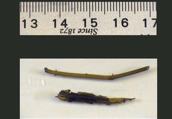
The child was given intravenous cefoxitin and taken to the operating room for exploration with a pre-operative diagnosis of a perforated appendicitis. Right lower quadrant exploration revealed a moderate amount of purulent fluid. The appendix was not inflamed. On exploration of the bowel a perforation was noted about 5 cm proximal to the ileocecal valve, with a piece of plant material partially extruding from a 2 mm perforation. The perforation was repaired, and an appendectomy was performed. The child’s postoperative course was uneventful. After completing a course of antibiotics he was discharged with a diagnosis of foreign body perforation of terminal ileum.
Pathology Report
Pathology reported an appendix measuring 5.9 cm in length and 0.5 cm in diameter with no evidence of acute appendicitis. The foreign body was reported as a plant stem about 0.1 cm in diameter (figure 1).
Retrospective review of the CT scan demonstrated a linear shadow adjacent to the ileum, consistent with the foreign body identified at surgery (figure 2).

2. PEM Stick on CT scan image
Discussion
Evaluation of abdominal pain in pre-adolescent children is often challenging, especially in infants and non-verbal young children. Differential diagnosis of abdominal pain in pre-adolescent children can frequently be simplified when localization can be determined by history and physical examination.
When pain and clinical exam localize pain to the right lower quadrant, appendicitis becomes the most important diagnosis to prove or rule out. Other acute surgical conditions that are associated with right lower quadrant abdominal pain in a toddler are torsion of the testis/ovary, bowel obstruction, intussusception and inflammation or perforation of Meckel’s diverticulum. Non-surgical conditions include ileitis, mesenteric lymphadenitis, viral illness, pneumonia, gastroenteritis, streptococcal pharyngitis, diabetes ketoacidosis, urinary tract infections or functional abdominal pain.
Appendicitis is a common concern in children who present to the emergency room for evaluation of abdominal pain. Typical age for the presentation of appendicitis in children is school age and adolescent.
Appendicitis Rare in Infants
Appendicitis is rare in infants and very young children with annual rate of 1 to 6 per 10,000 children between birth and four years [4,5] Newborn appendicitis can present as sepsis, and may be an indicator of other pathology, such as Hirschsprung’s disease. Complicated appendicitis, with perforation or phlegmon formation is more likely with young age, especially under five years of age.
Foreign body perforation of the small intestine is rare. The usual culprits include straight pins, toothpicks and broken orthodontic wires [1,3]. More recently, magnet ingestions have been reported to cause intestinal perforations [1].
Accurate history and careful physical examination are the key elements of evaluation of children with abdominal pain. Laboratory data may help to demonstrate the degree of illness, and cross-sectional imaging, such as CT scan and ultrasound, may aid in the diagnosis of appendicitis or other abdominal pathology. Ultrasound is now the preferred modality at most children’s hospitals for its rapidity and no risk of radiation exposure. Ultrasound use varies by institution policy. Ultrasound was not done in this case as the patient arrived past midnight and no ultrasound technician was available.
In this case, the CT imaging confirmed the presence of intra-abdominal pathology, but only in retrospect identified the true nature of the illness. The contrast use (IV vs. oral vs. rectal) varies by institution policy but several pediatric centers perform CT with IV only contrast for the evaluation of appendicitis.
Emergency physicians, surgeons and radiologists should consider foreign body perforation in the differential diagnosis of the acute abdomen in patients with a presentation atypical for other, more common, diseases.
REFERENCES
1. Magnetic foreign body injuries: a large pediatric hospital experience; Strickland M et al. Journal of Pediatrics 2014 Aug; 165(2):332-5.
2. A curious case of foreign body induced jejunal obstruction and perforation. Sarwa P et al. International Journal of Surgery Case Report 2014; 5(9): 617-619.
3. Cecal retention of a swallowed penny mimicking appendicitis in a healthy 2 year old. Pediatric Emergency Care 2004 Aug; 20(8):525-7.
4, Acute appendicitis in preschool-age children.
5.Sakellaris G, Tilemis S, Charissis G, Eur. J. Pediatr. – February 1, 2005; 164 (2); 80-3.
6. Acute appendicitis in children under 3 years of age. Diagnostic and therapeutic problems. Bagłaj M, Rysiakiewicz J, Rysiakiewicz K; Med Wieku Rozwoj – April 1, 2012; 16 (2); 154-61.


