A 40-year-old man is brought in by ambulance after sustaining a single stab wound to the left lower abdomen. The patient is asymptomatic with stable vitals, a negative FAST exam, no obvious evisceration and no clinical evidence of peritonitis on exam. On local wound exploration with the trauma surgery team, the wound violates the peritoneum.
With sensitivities and specificities quoted as high as 87% and 82% in select trauma patients and the highest sensitivity for hollow viscus injury, DPL remains a vital diagnostic tool when used in conjunction with advanced imaging.
Using tools commonly found in the ED and a DPL catheter, this relatively quick and safe procedure allows providers to access and evaluate the peritoneal cavity at the bedside and can be the deciding factor between laparotomy, observation or discharge.
Indications
The decision to perform a DPL should generally involve consultation with the trauma surgery team.
In general, in hemodynamically stable patients, DPL can be considered in the following:
- Blunt abdominal trauma where CT or FAST is not available or where imaging is equivocal
- Anterior abdominal stab wounds with violation of peritoneum on local wound exploration
- High suspicion for hollow viscus injury with negative or equivocal imaging
- Unreliable abdominal exam (i.e. altered mental status, intubated, spinal cord injury) with negative or equivocal imaging
- Changes in abdominal exam or vitals in observed patients with negative initial imaging
- Hemodynamically tenuous patients with blunt or penetrating trauma who cannot be safely transported out of the resuscitation bay (i.e. CT scanner, interventions for other injuries)
DPL should not be considered in patients who clearly warrant emergent laparotomy: hemodynamically unstable, gunshot wounds, stab wounds with evisceration or peritonitis – or in patients with positive diagnostic imaging (positive FAST or CT scan identifying an injury requiring surgical intervention). Other relative contraindications include: morbid obesity, pre-existing coagulopathy and cirrhosis/large volume ascites. Relative contraindications to the infraumbilical approach include: pregnancy greater than 12 weeks gestation, pelvic fractures and prior lower abdominal surgeries or scars.

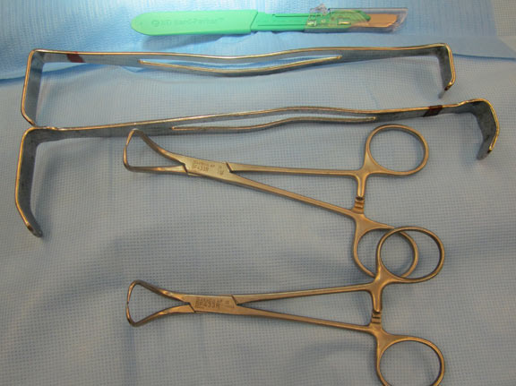
Equipment (for the open technique)
- Lidocaine 1% with Epi in 12cc syringe with injecting needle
- betadine or hibiclens
- fenestrated sterile drape
- scalpel (11 or 15 blade)
- retractors x 2
- towel clamps x 2
- DPL catheter
- 12 cc (or larger) syringe
- 1 liter bag of warm IV fluid (NS or LR) and IV tubing (without valve)
- 3-0 or 4-0 absorbable suture
- needle driver
Technique
There three different techniques for performing a DPL: open, semi-open and closed. The open technique was used on this patient so is described here.
A nasogastric tube and foley catheter must be placed and the stomach and bladder adequately decompressed. The patient must remain supine for the procedure. The entire procedure should be performed under sterile conditions.

01 The infraumbilical area is prepped and draped in sterile fashion. The area is then anesthetized using 1% lidocaine with epinephrine using wide local infiltration. Conscious sedation can be considered depending on urgency and the patient’s condition.

02 A 2-3 cm vertical midline skin incision is made approximately 2 cm below the umbilicus and the subcutaneous tissue is dissected down to expose the linea alba. Retractors can be used to hold back the wound edges and better expose the fascia.

03 The fascia on either side of the wound is then securely grasped using the towel clips and raised up away from the abdominal contents and a small vertical incision (~0.5 cm) is carefully made through the linea alba and fascia, opening the peritoneum.

04 The DPL catheter is then carefully advanced into the peritoneum through the fascial incision, directing it inferior (caudad) and posterior. Note that the goal to have the catheter tip resting anterior to the rectum (the most dependent portion of the peritoneum). The closed technique employs the Seldinger technique in which an introducer needle is passed through intact skin and fascia into the peritoneal cavity and the DPL catheter is positioned into the cavity over a guidewire. The semi-open technique is similar to the closed technique except that the skin is incised and the fascia exposed before the introducer needle is used to puncture the fascia into the peritoneum.

05 For lavage, the syringe is removed and a 1-liter bag of warm IV fluid (NS or LR) is connected to the DPL catheter using primed, sterile IV tubing. (The tubing should not have a 1-way valve as this prevents the return of fluid after lavage.) The IV fluid is allowed to flow into the peritoneal cavity (approximately 1 liter in adults, 10-15 ml/kg in children). If possible, the patient should be gently rocked from side to side as the fluid is infusing. Note that the IV bag should not be allowed to empty completely as this disrupts the siphon effect required to evacuate the effluent.
 Results interpretation
The DPL effluent should be sent for red blood cell count, white blood cell count and gram stain. A grossly positive result includes: immediate aspiration of 10 ml or greater of gross blood, aspirate containing gross enteric contents or vegetable matter. Although some disagreement exists, most studies quote a positive DPL result as one of the following criteria: red blood cell count > 100,000/mm3 or white blood cell count >500/mm3.
Some studies advocate checking lavage amylase and alkaline phosphatase, as they have higher specificity for small bowel injuries. Although, no consensus on cut-off values exists, > 19 IU/L and > 2 IU/L respectively has been suggested.
06 The catheter is connected to a 12 cc (or larger) syringe and aspirate obtained. If 10 ml of gross blood or enteric matter is obtained the DPL is considered grossly positive and the patient should be prepared for laparotomy. (Note that if < 10 ml of blood is obtained, the aspirate is returned to the peritoneal cavity before proceeding to lavage).
Case conclusion
Our patient had an asymptomatic anterior abdominal stab wound that violated the peritoneum and an equivocal CT scan and no contraindications to a DPL. In order to avoid a potentially harmful and unnecessary explorato
ry laparotomy in combination with the concern for a small hollow viscus injury, a DPL was performed. The DPL was not grossly positive on aspirate so a lavage was performed. The effluent analysis was negative and the patient remained asymptomatic. So after the catheter was removed and the wound repaired, the patient was discharged home and was continuing to do well on follow up several weeks later.

07 When nearly all of the fluid has infused, lower the bag to the ground and allow the effluent to drain out by gravity. At least 30% of the original infusate must be obtained for an adequate lavage sample. The effluent is sent for analysis; if negative, the catheter should be removed. The wound is irrigated and the fascia is closed with 1-2 simple interrupted absorbable sutures. Semi-open or closed technique does not require fascial closure. In all techniques the skin is closed using suture or skin staples.
Resources
- Schultz DJ, Weigelt JA: Diagnostic Peritoneal Lavage. In Operative Techniques in General Surgery Volume 5. Issue 3 Edited by: VanHeerden JA, Farley DR. Philadelphia, PA: WB Saunders; 2003:139-144.
- Sarin E, Kashuk JL, Cothren CC, et al. Diagnostic peritoneal lavage remains a valuable adjunct to modern imaging techniques. J Trauma. 2009;67:330-336.
- Biffl WL, Kaups KL, Cothren CC, et al. Management of patients with anterior abdominal stab wounds: a Western Trauma Association Multicenter trial. J Trauma. 2009;66:1294-1301.
- Jansen JO, Logie JR. Diagnostic peritoneal lavage—an obituary. Br JSurg. 2005;92:517–518.
- McAnena OJ, Marx JA, Moore EE. Contributions of peritoneal lavage enzyme determinations to the management of isolated hollow visceral abdominal injuries. Ann Emerg Med. 1991; 20:834-837.
Dr. Lina Tran is a 3nd year Emergency Medicine Resident at the Denver Health Emergency Medicine Residency Program. Dr. Peter Pryor is a faculty member at Denver Health, an Assistant Professor of Emergency Medicine at the University of Colorado School of Medicine and has an academic focus in medical photography.
1 Comment
Excellent review, but you lost me after these comments (I am loosely paraphrasing here): “in consultation with the trauma team,” “after you decompress the stomach with an NG tube,” “after you decompress the bladder (with a foley catheter).” Unless you are loaded with house staff or have a dedicated trauma team, I am afraid that the nursing resources required to get this done in a busy ED in a timely fashion is unrealistic. I would also argue that the ED charges (not costs) for a DPL are astronomical. Unless you are practicing in the 3rd world, DPL is dead.