edited by Mark Silverberg, MD
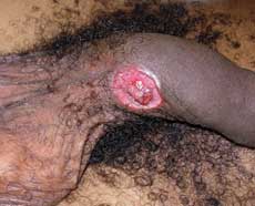
 On physical examination his vital signs are within normal limits and he is afebrile. He is well appearing, well nourished and in no acute distress. Upon examination of his genital region, a nontender, indurated, ulcerative lesion with raised edges about 1.5 cm in diameter is seen on the dorsum of his uncircumcised penis. There is no pus draining from the ulcer and no surrounding erythema or crepitus. Bilateral inguinal lymphadenopathy is present with mild tenderness on the left side. There is no scrotal swelling, crepitus or tenderness or expressible penile discharge. No other skin lesions or rashes exist on his body and there are no other areas of lymphadenopathy. The remainder of the physical examination is grossly unremarkable.
DISCUSSION:
The patient that presents with a single genital lesion can be a challenge to diagnose for the emergency physician. You have just met this gentleman and now you must discuss some of the most intimate details of his life. In an ideal situation you may have a full fifteen minutes to learn of his hygienic habits, sexual history and any number of other embarrassing details to determine the cause of his ailment and then you must embark upon a meticulous physical examination of a sensitive region.
The first questions which should come to mind are: Is this genital ulcer a skin manifestation of some other serious deep soft tissue infection and does it represent a serious life or limb threatening illness? One of the most sinister genital pathologies to consider is Fournier’s gangrene. Predispositions to this disease usually include diabetes, advanced HIV disease, malnutrition, alcoholism, chronic steroid use and extremes of age. Our patient’s history does not include any of these conditions. Furthermore, there are no signs on the physical examination that would indicate such a serious infection exists: He is well appearing and afebrile. He is non-tender and there is no crepitus in the region although subcutaneus emphysema is not seen in all cases of Fournier’s gangrene. From the given information, it can probably be concluded that if this ulcer is an infection, it is probably localized to the genital area with regional lymphadenopathy.
This lesion could feasibly represent a skin cancer, such as squamous cell carcinoma, but this is a very unlikely diagnosis.
After addressing the worse possible causative etiologies, we can safely move on to the more common causes of genital lesions which are usually due to sexually transmitted diseases. This differential diagnosis is vast but can be narrowed with a careful history identifying pertinent epidemiological factors and a detailed physical examination. Our patient is a young male with high risk sexual activity. Behavior such as this potentially exposes this individual to a number of different sexual transmitted diseases including HIV. Although not a cause of genital ulcers, co-infection with HIV can alter the presentation of certain skin lesions and impact treatment of many STDs.The most prevalent sexually transmitted diseases that cause penile lesions that will present in the emergency department are genital herpes and human papilloma virus (HPV). Primary genital herpes will present as numerous painful vesicles associated with constitutional symptoms such as fevers and myalgias. Our patient’s only complaint is a single painless ulcer, making the diagnosis of genital herpes unlikely unless it is very early in the course of the primary infection. If this patient has had previous exposure to HSV-1, a new infection with HSV-2 may be less severe and may present with isolated lesions, therefore herpes can be considered in the differential. HPV (subtypes 6 and 11) will cause condylomata acuminata which causes non-tender exophytic cauliflower-like lesions. Subtypes 16 and 18 can cause malignant lesions that may ulcerate as it invades local tissue, but such penile cancers are extremely rare in men who have been circumcised at birth and lesions are usually chronic and evolve over time.
This patient’s physical findings are classic for a primary syphilis infection, so we will have to investigate this entity more thoroughly through laboratory testing. Though only 9,000 cases were reported to the CDC in 2007, it is far more common in the United States compared to the other etiologic agents to be discussed on the differential. Lymphogranuloma venereum (LGV), chancroid and granulomatosis inguinale all present with ulcerative lesions but are rarely seen in the United States. Lymphogranuloma venereum caused by Chlamydia subtypes L1, L2, and L3 begins with a painless ulcer. As the disease progresses, painful lymphadenopathy and bubo formation occur and is associated with systemic constitutional symptoms. Ultimately the disease may progress to proctocolitis. Though prevalence in the United States is low, the incidence in Western Europe has risen over the past few years in men who have sex with men making it an important disease entity for EPs to become familiar with.
Another penile lesion to consider is chancroid. It usually presents as a painful ulceration caused by H. ducreyi and also leads to lymphadenopathy and bubo formation. However, only 27 confirmed cases were reported in the United States in 2007. His lesion is painless making this diagnostic entity unlikely. The genital lesions in granulmatosis inguinale may be painless and also present as ulcerations that are beefy red and bleed easily on contact. These diseases should be considered especially if your ED is providing for a large immigrant community.
Cutaneous fungal infections can cause single focal lesions or diffuse balanitis and are another possible consideration in the differential diagnosis. These infections typically occur in uncircumcised men and are quite itchy but usually painless. Most people with these infections end up having diabetes. Our patient’s lesions are not characteristic for a fungal lesion and he does not have any history of diabetes so this diagnosis is again unlikely.
Other conditions that should be considered are noninfectious causes of genital lesions. Bechet’s disease is an autoimmune disorder characterized by recurrent mucosal membrane ulcers and uveitis in addition to genital ulcers which can be seen in up to 50% of patients with this autoimmune disease. Again, this patient’s only physical finding is a solitary ulcer with no signs of a systemic disease making this diagnosis unlikely. Pearly penile papules may also be considered. They present as small round lesions that are arranged circumferentially in one or several rows usually located on the corona of the glans penis. This condition is often misdiagnosed as a sexually transmitted disease but the cause is idiopathic. Again, the location and description of this patient’s lesion does not suggest this diagnosis.
Other questions that need to be asked during the history, which may be quite personal, are hygienic habits and sexual practices. Traumatic lesions can ulcerate if not properly attended to. Pressure ulcers can result from jewelry or other accessories if worn too tightly or repeatedly rubbed on the genitals. Perhaps he injured himself while shaving the area and has developed a small wound infection. Maybe there was some other incident which he is too embarrassed to admit to. Second and third degree burns from hot wax are not unheard of and can also ulcerate if super-infected. However, these traumatic lesions would most likely cause pain, which is not present in our patient.
Allergic reactions are another consideration in this case. Lotions or creams or latex in condoms can result in local irritation/lesions which can also become secondarily ulcerated and possibly infected. An insect bite to the area is another possibility although our current history does not support any of these diagnoses.
With a broad differential diagnosis in place, we can now turn our attention to the laboratory work up for our patient which should include a CBC, RPR, serology for HSV and H. ducreyi culture, gram stain of the lesion and if available, a tzanck smear and dark field microscopy. There is no definitive lab test readily available that can distinguish between the subtypes of Chlamydia that cause lymphogranuloma verenum, so diagnosis of this disease is usually clinical. For this reason, it must be seriously considered if the rest of the workup is negative. He should also be offered a HIV screening examination. Dependent on the laboratory capability, these test results may not be available in a timely fashion. Since this patient is stable, it is reasonable to discharge him with a plan to follow up these lab results once sent.
Two days later it is reported that this patient’s RPR is positive and he is called back to the clinic for re-evaluation. RPR is a screening test used to detect nonspecific antibodies that may indicate the presence of Treponema pallidum, the causative agent of syphilis. A more sensitive and specific diagnostic test such as FTA-ABS should now be drawn to confirm this diagnosis, but early in the disease process the test for specific antibodies to syphilis may still be nonreactive. The sensitivity of the FTA-ABS for primary syphilis is only 50-85%, so even if his FTA test is negative, there is still a high enough likelihood of T. pallidum infection for this patient that he should undergo treatment for syphilis at this time.
Though penicillin has been well established as the treatment for syphilis, a well-controlled, prospective study to determine the optimal dose or duration of therapy has never been performed. Current recommendations and guidelines for treatment are based on extrapolation of older data and limited clinical experience. Treponema pallidum is isolated in the CSF in about one-third of patients who present with early primary syphilis, indicating a propensity for spirochetes to invade the CNS. However, it has been shown that benzathine penicillin does not achieve adequate levels in the CSF. With this in mind, physicians should be aware of the possibility of treatment failures and progression of the neurological manifestations of this disease. Therefore it is essential in the patient’s management to provide education and proper long term follow up.
This patient has no known drug allergies and is treated with 2.4 million units of benzathine penicillin given intramuscularly once. Alternative therapies include doxycycline 100mg bid for 15 days, tetracycline 500mg qid for 15 days or azithromycin 2 grams once, although there is recent concern for the increase in resistance to azithromycin. It is important to remember to inform this patient of the possibility of the Jarisch-Herxheimer reaction. This reaction resembles a gram negative sepsis with abrupt onset of fevers, myalgias, chills and hypotension with various degrees of severity. It typically develops a few hours after antibiotics are initiated due to the torrent of immune modulators that are released as the spirochetes are destroyed. This syndrome usually resolves within a day or so and it occurs in approximately 50% of those treated for primary syphilis. After our patient receives his antibiotics, he is advised to follow up to repeat the quantitative RPR at 3, 6 and 12 months. If he is adequately treated, the RPR should return to negative. A positive RPR after one year may represent treatment failure or reinfection.
DIAGNOSIS: Primary Syphilis