A 35-year-old female with a previously diagnosed ectopic pregnancy, status post methotrexate therapy presents to the ED for near syncope and rectal pain. She states that she usually has irregular periods, but thinks she was four weeks pregnant when she received her first dose of methotrexate 10 days ago. Three days ago, she received a second dose because her beta-HCG had not started to decrease. She is in the ED now for multiple near syncopal episodes today occurring only when she stands up from a lying position and also a feeling of rectal pressure that is not relieved by bowel movements. She denies any complete loss of consciousness, chest pain, SOB, palpitations or abdominal pain and states that once she is standing for a while she is no longer light-headed and can walk around just fine. She has been having minor vaginal bleeding, but that is unchanged from when her ectopic was originally diagnosed a week and a half ago.
On physical exam, her pulse and temperature are normal. Her BP is soft at 103/62 but she is thin and walks to her treatment room from the waiting room with no apparent symptoms or difficulty. Her abdomen is benign and the rest of the exam is normal, but you decide to skip the rectal exam. Instead, you do a quick bedside point of care ultrasound (POCUS). Of course, you also order labs and IV fluids. One of the images you obtain on POCUS is shown below.
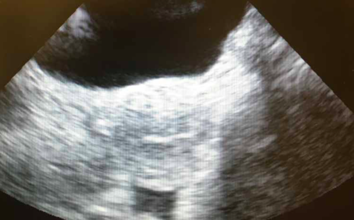
 What does the image demonstrate? Based on this image, what should be your next move?
The image is a transabdominal view of the urinary bladder and the uterus. Whenever I have concern for a ruptured ectopic, as I did in this case due to near syncope and tenesmus, the first ultrasound image I usually take is of Morrison’s pouch. That was negative in this case. The next image I usually take is of the pelvis. In this case, there is a small amount of free fluid (think blood) behind the uterus. (See image below with labels: EMS = endometrial stripe, FF = free fluid). This blood is in the rectouterine pouch, AKA pouch of Douglas, AKA cul-de-sac. Fluid here may produce tenesmus by it’s irritating effect on the rectum.

Your next move should probably be a call to the patient’s obstetrician. If the patient were to become unstable, she would need to go directly to the OR, but if she remains stable, as she did in this case, the obstetrician will have to decide between immediate operative management or observation with serial vitals and hemoglobins. In this case, I figured with methotrexate failure the obstetrician would want to go to the OR, but he was not convinced that the amount of fluid I had documented was significant, so he requested a formal ultrasound. The formal study was read out as showing, “a small amount of free fluid in the cul-de-sac”. The obstetrician decided to admit for observation and the patient remained stable, but by the next morning the hemoglobin had dropped two grams and the repeat ultrasound showed a “moderate” amount of free fluid. The patient was rushed to the OR and fortunately did well. I still don’t understand why the patient wasn’t taken to the OR on the first day. To me it was an obvious ruptured ectopic. Why not take her to the OR while she is stable?
Pearls + Pitfalls
- Assess Stability: Try to avoid sending a potentially unstable patient to radiology. If you have the skills, perform a bedside ultrasound yourself and call your surgeon early. If not, have your ultrasound tech come to the ED to scan at the bedside.
- Basics: The EM physician rarely needs to do a trans-vaginal scan. With a trans-abdominal you can usually tell if there is an IUP or not (to rule out ectopic), and if there is free fluid or not (to rule out rupture). This, and evaluation for cardiac motion should be the extent of a limited bedside ultrasound. The order of appearance of structures within the uterus in pregnancy is: gestational sac (beware of pseudosacs) then a yolk sac, then a fetal pole, then finally cardiac motion.
- Free Fluid: Free fluid in the pelvis or abdomen should be considered blood until proven otherwise. Blood and ascitic fluid may look the same on ultrasound. In the proper clinical scenario, such a hemodynamically stable patient with no history of trauma and a benign exam, ascites may be considered. Other causes of free fluid may include pus or bowel or bladder rupture.
- Start at Morison’s: Begin by scanning the hepatorenal recess (Morison’s pouch). This is the most dependent position in a supine patient so free fluid may collect here. If you see fluid here, call for help ASAP. Any patient with a positive pregnancy test and free fluid visible on bedside ultrasound should be suspected of having a RUPTURED ectopic pregnancy until proven otherwise.
- The Pelvis: After Morrison’s pouch evaluate the pelvis. This is the most dependent part of the abdomen when upright. To get the best views, the patient should ideally have a full bladder to serve as an acoustic window. If she just gave a urine sample, you will have to do your best. Look for hypoechoic free fluid in the vesicouterine and rectouterine spaces and assess the uterus for the presence or absence of an IUP. In a ruptured ectopic all you may see is a confusing mess of heterogeneous clotted blood that can even make it hard to delineate the uterus. If you have done plenty of scans of the normal pelvis, which you should have, you will be able to tell that “something looks wrong” though you may have a hard time accurately determining exactly what you are looking at.
- Beware: A cornual ectopic, which is the most dangerous type of ectopic due to rich blood supply to the cornua, can be close enough to the uterus to fool you into thinking it’s an IUP if you are not careful. Make sure there is at least a 2-cm mantle of myometrium on all sides of a gestational sac before diagnosing and IUP. Also, beware of pseudosacs, irregular intrauterine sacs that can co-occur with an ectopic.