
There are times when you need to perform an ultrasound under sterile conditions. Do you know how?
“A bet’s a bet…” you chirp at your resident as you grab the bag of chocolate covered raisins out of her hand. Your resident may have lost her snack food for the evening, but she can’t hide her pleasure in knowing that your “psychic” ability to predict a patient’s hematocrit of 18% means she will be getting to do a central line tonight. Before you can even begin enjoying the spoils of your win, your resident has taken off to grab a central line kit and the ultrasound machine.You stand at the patient’s bedside, watching one of your star pupils localize the IJ with bedside ultrasound. As she pushes the ultrasound machine away and begins prepping the neck, you casually mention “Why don’t we do this one in a dynamic fashion under real-time ultrasound guidance”. Without blinking, your resident answers “Sure. Sounds like a great idea. Before you start eating those chocolate raisins, double or nothing says I can get this line in under 3 minutes.” You pause for dramatic effect, and reply back “I’ll bring your shift snacks for a month if you can place this central line in a sterile fashion using only your two hands and the ultrasound machine”. She counters, “You’ve got yourself a bet.”
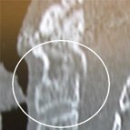
After prepping and draping the patient’s neck, your ultrasound guru-in-training squirts a glob of gel on the linear transducer while it’s still sitting upright in its slot next to the machine. You’re thankful that your patient is sedated and intubated as your resident decides to use the patient’s abdomen as the equipment table. Opening up the prepackaged sterile kit with a flourish, she quickly lays out the sterile drape, the sterile probe cover, and the sterile ultrasound gel. After donning her sterile gloves, your resident tunnels her hand into the probe cover and carefully grabs the gel-coated transducer with the sterile sleeve. With her other gloved hand, she skillfully maneuvers the probe cover down along the transducer cord without violating her sterile field
 (top).
Grabbing the sterile prepackaged ultrasound gel she tops off the covered transducer prior to beginning her scan for the patient’s internal jugular vein (bottom). With 30 seconds to spare, she localizes the vein, inserts a double lumen catheter via the Seldinger technique, and gently secures her beautifully executed central line with suture and a Tegaderm. As she disposes of her own sharps, you can’t help but marvel at how facile your residents are with ultrasound these days.
You happily hand back over your coveted chocolate covered raisins, and casually ask her how her international elective went last month. With a winning smile she answers, “Oh, I forgot to mention, I had to postpone my away elective for a couple of months. Since I want to stay and do an ultrasound fellowship, I did an ultrasound elective instead…. We did enough guided central lines that I could have probably done that last one with my eyes closed…” With a wink she echoes, “Like you’ve always said, it’s nice to have help, but it’s best to learn how to do things on your own. Now hand over the chocolate Dr. Wu”
After prepping and draping the patient’s neck, your ultrasound guru-in-training squirts a glob of gel on the linear transducer while it’s still sitting upright in its slot next to the machine. You’re thankful that your patient is sedated and intubated as your resident decides to use the patient’s abdomen as the equipment table. Opening up the prepackaged sterile kit with a flourish, she quickly lays out the sterile drape, the sterile probe cover, and the sterile ultrasound gel. After donning her sterile gloves, your resident tunnels her hand into the probe cover and carefully grabs the gel-coated transducer with the sterile sleeve. With her other gloved hand, she skillfully maneuvers the probe cover down along the transducer cord without violating her sterile field
(top).Grabbing the sterile prepackaged ultrasound gel she tops off the covered transducer prior to beginning her scan for the patient’s internal jugular vein (bottom). With 30 seconds to spare, she localizes the vein, inserts a double lumen catheter via the Seldinger technique, and gently secures her beautifully executed central line with suture and a Tegaderm. As she disposes of her own sharps, you can’t help but marvel at how facile your residents are with ultrasound these days.
You happily hand back over your coveted chocolate covered raisins, and casually ask her how her international elective went last month. With a winning smile she answers, “Oh, I forgot to mention, I had to postpone my away elective for a couple of months. Since I want to stay and do an ultrasound fellowship, I did an ultrasound elective instead…. We did enough guided central lines that I could have probably done that last one with my eyes closed…” With a wink she echoes, “Like you’ve always said, it’s nice to have help, but it’s best to learn how to do things on your own. Now hand over the chocolate Dr. Wu”
Brady Pregerson, MD, oversees QI for ED Ultrasound at Cedars-Sinai Medical Center in Los Angeles. For more images, check out Real-Time Readings at www.epmonthly.com.
Teresa Wu, MD, a clinical assistant professor in EM at Florida State University, completed her ultrasound fellowship at Stanford University Medical Center.
Teresa Wu, MD, a clinical assistant professor in EM at Florida State University, completed her ultrasound fellowship at Stanford University Medical Center.
For 10 steps for performing the sterile ultrasound-guided procedure, see next page
{mospagebreak}
10 Steps for Performing a Sterile Ultrasound-Guided Procedure
1. It is very helpful to have an extra set of hands available for most sterile ultrasound guided procedures, but it can also be done as a sole operator with a bit of extra preparation and planning.
2. Prep and drape the target area on the patient first.
3. Prime the face of the ultrasound probe with gel and position the probe so that it is readily accessible.
4. Open up and lay out the sterile ultrasound sheath (also referred to as a probe cover) and sterile ultrasound gel. If your department does not have sterile ultrasound kits available, open up a set of large sterile gloves and squeeze a small amount of sterile ultrasound gel onto the glove without violating the sterile field. Also, make a mental note to remind your department chair that in 2007, and ED with bedside ultrasound should have sterile probe covers as well.
5. Put on a facemask, sterile gown, and gloves in the traditional fashion.
6. Tunnel your gloved hand into the transducer sheath and grab the ultrasound probe with the sterile sheath.
7. If you have an extra set of hands available, have your assistant prime the transducer with ultrasound gel and position the probe for you to grab with the sterile sheath. (left – imagine blue glove is from assistant)8. Use your other sterile hand to slide the ultrasound sheath down over the transducer cord. Consider using a hemostat to clip the probe cover to the wide field sterile barrier as this may help minimize the risk of the probe falling to the floor once you put it down. This risk is increased by the probe sheath, which tends to have a low coefficient of friction.
9. Attempt to remove any air bubbles that may have accumulated in the gel between the transducer and the sterile sheath.
10. Finally, apply a copious amount of sterile gel over the sheath-encompassed transducer and begin your scan.
Brady Pregerson, MD, oversees QI for ED Ultrasound at Cedars-Sinai Medical Center in Los Angeles. For more images, check out Real-Time Readings at www.epmonthly.com.
Teresa Wu, MD, a clinical assistant professor in EM at Florida State University, completed her ultrasound fellowship at Stanford University Medical Center.
Teresa Wu, MD, a clinical assistant professor in EM at Florida State University, completed her ultrasound fellowship at Stanford University Medical Center.